Download
1 / 33
330 likes | 431 Vues
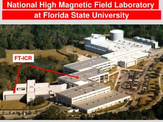
Florida State University, National High Magnetic Fields Laboratory. Conformational Changes Associated with Muscle Activation and Force Generation by Pulsed EPR Methods. Piotr Fajer. myosin head. Motor proteins. Ca activation. actin. troponin C. myosin. Force generation.

E N D
Florida State University, National High Magnetic Fields Laboratory Conformational Changes Associated with Muscle Activation and Force Generation by Pulsed EPR Methods Piotr Fajer
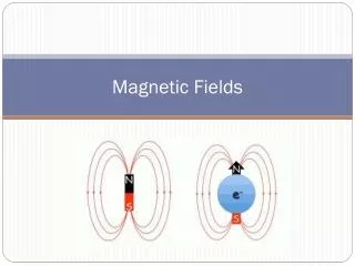
myosin head Motor proteins Ca activation actin troponin C myosin Force generation function demands large conformational changes;
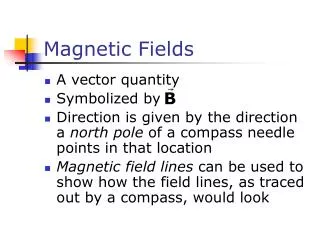
Why EPR ? • Orientation • Dynamics • Distances • 2o structure
O O N O InVSL cysteine O cysteine N O H N O N N O O IASL MSL Labeling Cysteine Scanning Cysteine scanning Native cysteines
nitroxide - nitroxide Dipolar EPR: distances Rabenstein & Shin, PNAS, 92 (1995) Non-interacting spins Double labeled sensitivity: 8-20 Å
Echo /2 Nitroxide (ms) Dipolar interaction Gd3+ (s) Distance : metal-nitroxide Pulsed EPR T1 echo intensity Time Sensitivity: 10–50 Å
DEER(Double Electron Electron Resonance) Echo /2 Dipolar interaction nobserve t2 t1 t1 npump t Echo Modulation Milov, Jeschke Model spectra Long Distance: 18 –50 Å Sensitive to distance distribution 25 Å 38 Å
Dipolar EPR myosin cleft closure myosin head interactions in smooth muscle troponin opening of K+ - channel Site specific spin labelling structure of troponin I Applications
Actin binding cleft conformation 416 537 A. Málnási-Csizmadia, C. Bagshaw, P. Connibear force Cleft closure associated with lever swing
EPR distances • distribution of distances • changing fraction of each • equilibrium of CLOSED and OPEN states shifts towards CLOSED in the presence of actin
Smooth muscle regulation RLC- RLC MD-MD Wendt et al. (1999) Wahlstrom et al. (2003) Taylor Cremo • Hypothesis: heads stick together inhibiting ATPase
RLC single cysteine mutants Taylor Cremo
EPR distances • The measured distances are consistent with the Taylor model • The N-terminal portion is further apart than either model
Troponin: Collapse of central helix 47 Å 37 Å Vassylyev et al. PNAS, 1998 Tung et. al, Protein Sci, 2000
Ca switch mechanism shown in isolated TnC but NOT in ternary complex of TnI, TnC and TnT Questions: what is the structure of TnC in ICT complex ? what are the Ca induced conformational changes in ICT ? Troponin
Collapse of TnC central helix Spin labels: 12, 51, 89, 94 Gd3+: sites III & IV
Isolated TnC Excellent agreement with X-ray and NMR TnC in solution is extended
Ternary complex 37 Å N- to C-domain distance decreases by 9 Ǻ central helix bends in a complex
15-94 15-136 12-136 N-domain: Homology model for Ca2+ switch in troponin (assume no changes in the N-domain which senses Ca) • distances consistent with the TnC based homology model
C-domain of TnC TnI 51 TnC 100 All distances are in (Ǻ) TnI N-terminal helix moves v. little (2Å) with respect to TnC C-domain on Ca2+ binding.
TnC is more compact in ternary complex than isolated TnC. Calcium switch might well be same in troponin complex as in isolated TnC. 3. N-domain of TnI remains in proximity of C-domain of TnC. N domain movement central helix bending Tn (+ Ca) Tn (- Ca) TnC + = Conformational changes in a complex
Opening of K+ channel Y. Li, E. Perozo Closed (x-ray) Open (homology) Homology model is wrong.
Fidelity of the EPR distances Scatter = 6 Å
Molecular Dynamics Distance Spin-spin angle
EPR v. X-ray/MD-MC Modelling the spin label decreases scatter = 3 Å
Site Directed Spin Labeling EPR Hubbell, 1989 “cysteine scanning” from 130-146
P1/2(O2)/ P1/2(CROX) P1/2(O2)/ P1/2(CROX) Secondary structure determination 4 P1/2= 60 mW amplitude 2 P1/2= 20 mW 0 0 5 10 15 power ½ (mW) ½
Computational models TnI inhibitory region -helix (x-ray) X-ray CS data, homology model Vassylyev et al PNAS 95:4847 ‘98 -hairpin loop (nmr) Neutron scattering Tung et al Prot.Sci. 9:1312 ‘00
Identifying the interface between subunits Binary/ternary “difference” map 130 131 132 130-136 TnT imprint 133 134 135 136 200 137 tICT/tIC Ternary: TnI mutants 0.006
Dipolar EPR excellent for 10-20 A Pulsed EPR extends the range to 20-50 A “Easy” protein chemistry Large macromolecular complexes Determination of secondary structure. Summary
Hua Liang Song Likai Clement Rouviere Louise Brown Ken Sale The Lab • Collaborators • Clive Bagshaw ~ U. Leicester • Málnási-Csizmadia ~ Eötvös U. • E. Perozo ~ U. Virginia