Download
1 / 55
780 likes | 1.16k Vues
ADRENAL DISORDERS. Hasan AYDIN, MD Yeditepe University Medical Faculty Department of Endocrinology and Metabolism. Histologic Z ones of A drenal C ortex. Zona Glomerulosa Aldosterone Zona Fasiculata Cortisol and androgens Zona Reticularis Cortisol and androgens.

E N D
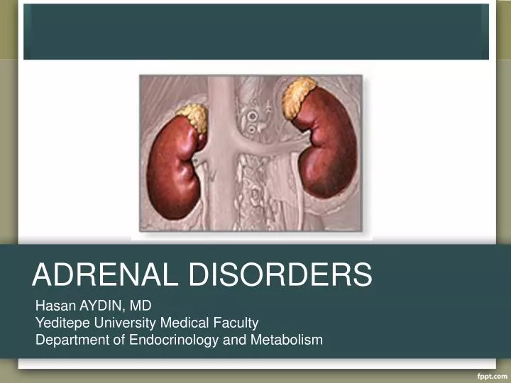
ADRENAL DISORDERS Hasan AYDIN, MD Yeditepe University Medical Faculty Department of Endocrinology and Metabolism
Histologic Zones of Adrenal Cortex • Zona GlomerulosaAldosterone • Zona FasiculataCortisol and androgens • Zona ReticularisCortisol and androgens
Diseases of Adrenal Gland • Diseses of Adrenal Cortex • Cushing syndrome • Adrenal failure • Congenital adrenal hyperplasia • Diseases of Adrenal Medulla • Pheochromocytoma • Hyperaldosteronism (Conn’s syndrome)
Cushing’s Sydrome vs. Disease • Chronic glucocorticoid excess, whatever it’s cause • Cushing’s diasease: a spesific type of Cushing’s sydrome due to excessive pituitary ACTH secretion from a pituitary tumor.
Cushing’s Syndrome • ACTH dependent • Pituitary adenoma (Cushing’s Disease) • Nonpituitary neoplasm (ectopic ACTH) • ACTH independent • Iatrogenic (glucocorticoid,megestrol acetate) • Adrenal neoplasm (adenoma,carcinoma) • Nodular adrenal hyperplasia primary pigmented nodular adrenal disease massive macronodular adrenonodular hyperplasia food-dependent (GIP-mediated )
Cushing’s Disease • 70 % of cases of Cushing’s sydrome • Female/male 8:1 • Age at diagnosis : childhood –70 years Etiology: Corticotroph-cell pituitary adenomas Corticotroph cell hyperplasia Tumors are typically smaller than 10 mm
Ectopic ACTH Hypersecretion • 15-20 % of patients with ACTH dependent Cushing’s Sydrome • Tumors causing the ectopic ACTH syndrome • Small cell carcinoma of the lung (50% of cases) • Pancreatic islet tumors • Carcinoid tumors (lung,thymus,gut,pancreas,ovary) • Medullary carcinoma of the thyroid • Pheochromocytoma
Adrenal Cushing’s Syndrome • Adrenal neoplasm (adenoma,carcinoma) • Nodular adrenal hyperplasia • Primary pigmented nodular adrenal disease • Massive macronodular adrenonodular hyperplasia • Food-dependent (GIP-mediated )
Diagnosis • Urine free cortisol in 24 hour urine samples – 80-100 mg/24 h is normal • Used in screening
Diagnosis • Absence of diurnal rhythm is the hallmark of diagnosis Serum cortisol levels exceeding 7 mg/dL at midnight indicates absence of diurnal rhythm
Diagnosis • Dexametasone supression test Low dose –for screening 1 mg dexametasone at bedtime (23 00hour) Determine plasma cortisol early following morning Plasma cortisol < 1.8 mcg/dl - normal Dexamethasone 4 x 0.5 mg for two days 17 hydroxycorticosteroid excretion greater than 4 mg/24 h on the second day of dx administration= Cushing syndrome
Differential Diagnosis • Plasma ACTH: Differentiate ACTH dependent and non dependent forms Patients with ACTH –secreting neoplasms usually have plasma ACTH levels > 10 pg/ml frequently greater than 52 pg/ml. • Pituitary MRI ACTH-dependent patients adenoma on MRI likehood of cushing disease 98-99% Incidentaloma 10%
Inferior Petrosal Sinus Sampling • Distinguishing pituitary from non pituitary ACTH dependent Cushing’s syndrome • Simultaneous inferior petrosal sinus and peripheral ACTH measurements before and after CRH stimulation • IPS/P > 2 pituitary ACTH secreting tumor • IPS/P < 1.8 ectopic ACTH • Diagnostic accuracy 100% in the differential diagnosis of ACTH dependent Cushing’s syndrome
Treatment Remove or destroy the basic lesion Correct the hypersecretion of adrenal hormones • microsurgery • radiation therapy • pharmocologic inhibition of ACTH secretion ketoconasole,metyrapone,amimoglutethimide, mitotane
Primary adrenal insufficiency Autoimmune (70%of patients) Infections Tuberculosis (20% of patients) menincococcus,pnumococcus, fungal,HIV Medications (ketoconasole, dilantin,phenobarbital,rifampin, etomidate, metyrapone) Malignancy (Primary,metastatic) Adrenal hemorrhage (spontan,traumatic, coagulopathy/heparin/coumadin) Familial Infiltrative diseases (amiloidosis,sarcoidosis, hemochromatosis) Secondary adrenal insufficiency Exogenous glucocorticoid withdawal Following cure of cushing’s sydrome Hypothalamic or pituitary disease (Tumor,sarcoidosis,hemorrhage, autoimmune,postoperative) Isolated adrenocorticotropic hormone deficiency Causes of Adrenal Insufficiency
Presentation Highly variable Duration of disease Whether deficiency is primary or secondary
Glucocorticoid deficiency Cardiovascular hypotension Gastrointestinal anorexia,nausea,weight loss, abdominal pain, diarrhea Cutaneous hyperpigmentation (secondary to ACTH precursors) vitiligo(secondary to autoimmune disease) Muscular fatigue,weakness,malasie Neurologic confusion,apathy,lethargy,psycosis Mineralocorticoid Deficiency Cardiovascular hypovolemia, decreased cardiac output, Potential Clinical Manifestations
Acute Adrenal Crisis occurs in patients with Addison’s disease who are exposed to the stress of infection, trauma,surgery or dehydration
Clinical Features of Acute Adrenal Crisis • Hypotension and shock • Fever • Dehydration,volume depletion • Nausea,vomiting,anorexia • Weakness, apathy, depressed mentation • Hypoglycemia
Glucocorticoid deficiency Lymphopenia Eosinophilia Hypoglycemia Anemia Minerolocorticoid deficiency Hyponatremia Hyperkalamia Acidosis Azotemia Hypercalcemia Laboratory Findings of Adrenal Insufficiency
Diagnosis of Adrenocortical Insufficiency Since basal levels of adrenocortical steroids in either urine or plasma may be normal in partial adrenal insufficiency, tests of adrenal cortical reserve are necesseary to establish the diagnosis Cortisol > 20 mg/day at any time of day - diagnosis very unlikely Hemodynamic instability - cortisol < 20 mg/day - suspicious
ACTH Stimulation Test Performed at any time of day A baseline cortisol sample is obtained and 250 mg synthetic ACTH (cosyntropin) is then administered intravenously. Cortisol samples are drawn 30 and 60 min later. Plasma cortisol >18 mcg/dl excludes the diagnosis
Plasma ACTH Level • Used to differentiate primary and secondary forms Secondary adrenal insufficiency plasma ACTH <30 pg/mL ( 7pmol/L) Primary adrenal insufficiency plasma ACTH >52 pg/mL
Treatment ofAcute Adrenal Crisis • Glucocorticoid replacement Administer cortisol (hydrocortisone 100 mg i.v. Every 6 hours for 24 hours When patient stable reduce dosage to 50 mg every 6 hours Taper the maintenance therapy by day 4 or 5 and add minerolocorticoid therapy as required Maintain or increase the dose to 200-400 mg/day if complications persist or occur • General and supportif measures Correct volume depletion,dehydration and hypoglycemia With i.v. saline and glucose Evaluate and correct infection and other precipitating factors
Maintenance Therapy • Cortisol 15-20 mg in AM. and 10 mg at 4-5 PM • Fludrocortisone, 0.05-0.1 mg orally in AM • Clinical follow-up : maintenance of normal weight, blood pressure, and electrolytes • Patient education, identification card or bracelet • Increase cortisol dosage during stress
Adrenal Hormone Synthesis Cholesterol Mineralocorticoids Glucocorticoids Sex hormones Cholesteol desmolaz 17α-OH 17,20-liyaz 17-OH pregnenolon DHEA Pregnenolon 3β-HSD 3β-HSD 3β-HSD 17α-OH 17,20-liyaz Δ-Androsteodione Progesteron 17-OH progesteron 17β-HSD 21-OH 21-OH 11-Deoxycorticosterone 11-Deoxycortisol Testosterone 5α-redüktaz tip2 11β-OH Aromataz 11β-OH Corticosterone Cortisol Dihydrotestosterone Estradiol 18-OH 18-OH B 18-HSD Aldosterone
Treatment • Corticosteroid replacement • Mineralocorticoid replacement
Hyperaldosteronism • Primary • Adrenal adenoma • Adrenal carcinoma • Bilateral hyperplasia • Secondary • Renal artery stenosis • Edematous states (cirrhosis, renal failure)
Hyperaldosteronism • ↑BP + K+↓ or low normal K+ • ↑BP in young person • ↑BP difficult to control
Signs and Labs • Hypertension • Hypokalemia • Urine potassium wasting • Increased aldosterone • Low renin
Hyperaldosteronism, Diagnosis • Serum K < 3.6 mEq/L • Plasma renin activity (PRA) < 1 ng/ml • Plasma aldosterone >22 ng/dL • Urine aldosterone > 14 mcg/24hrs • Urine K > 40 mEq/24 hrs • Plasma aldosterone:PRA ratio > 50:1
Hyperaldosteronism, Diagnosis • CT scan • Adrenal vein sampling
Hyperaldosteronism, Treatment • Bilateral hyperplasia-medical, spironolactone, amiloride. • Unilateral adenoma- adrenalectomy.
Catecholamine Producing Tumors Neural Crest Neuroblastoma Sympathoadrenal Progenitor Cell (Neuroblasts) Chromaffin Cell Sympathetic Ganglion Cell Intra-adrenal Extra-adrenal Pheochromocytoma Ganglioneuroma
Pheochromocytoma • 0.01-0.1% of HTN population • Found in 0.5% of those screened • M = F • 3rd to 5th decades of life • Rare, investigate only if clinically suspicion: • Signs or Symptoms • Severe HTN, HTN crisis • Refractory HTN (> 3 drugs) • HTN present @ age < 20 or > 50 ? • Adrenal lesion found on imaging (ex. Incidentaloma)
Pheo: Signs&Symptoms • The five P’s: • Pressure (HTN) 90% • Pain (Headache) 80% • Perspiration 71% • Palpitation 64% • Pallor 42% • Paroxysms (the sixth P!) • The Classical Triad: • Pain (Headache), Perspiration, Palpitations • Lack of all 3 virtually excluded diagnosis of pheo in a series of > 21,0000 patients
Localization: Imaging • CT abdomen • Adrenal pheo SEN 93-100% • Extra-adrenal pheo SEN 90% • MRI • > SEN than CT for extra-adrenal pheo • MIBG Scan • SEN 77-90% SPEC 95-100%
Pheochromocytoma, Treatment • Treatment is surgery • Must medically optimize prior to surgery • Treat HTN • Expand intravascular volume • Control cardiac arrhythmias
Preop Preperation Regimens • Combined α + β blockade • Phenoxybenzamine • Selective α1-blocker (ex. Prazosin) • Propanolol • Metyrosine • Calcium Channel Blocker (CCB) • Nicardipine
Incidentaloma • Found on work up for another cause, not on cancer workup • US 0.1% • CT 0.4 to 4.4% • MRI • 70-94% are benign and nonfunctional • >3cm more likely to be functional • Up to 20% may be subclinically active • Increase with age, no change in sex • 5-25% will increase in size by at least 1cm