Download
1 / 58
740 likes | 1.3k Vues
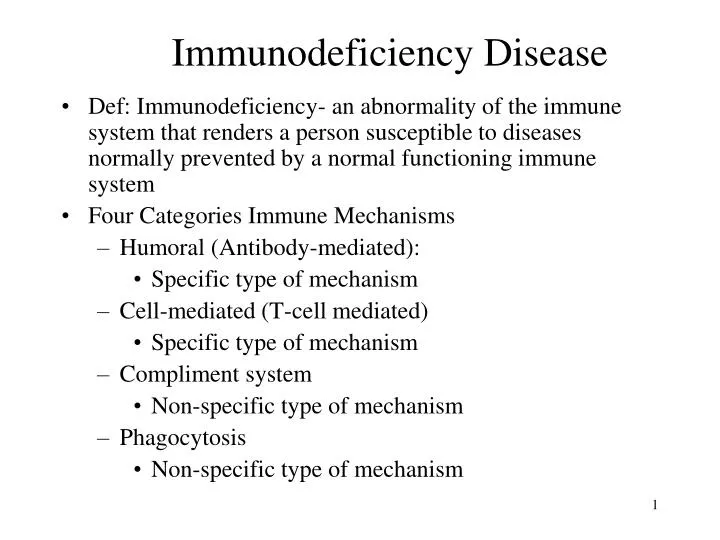
Immunodeficiency Disease. Def: Immunodeficiency- an abnormality of the immune system that renders a person susceptible to diseases normally prevented by a normal functioning immune system Four Categories Immune Mechanisms Humoral (Antibody-mediated): Specific type of mechanism

E N D
Immunodeficiency Disease • Def: Immunodeficiency- an abnormality of the immune system that renders a person susceptible to diseases normally prevented by a normal functioning immune system • Four Categories Immune Mechanisms • Humoral (Antibody-mediated): • Specific type of mechanism • Cell-mediated (T-cell mediated) • Specific type of mechanism • Compliment system • Non-specific type of mechanism • Phagocytosis • Non-specific type of mechanism
Immunoglobulins • An antibody or immunoglobulin: • Large Y-shaped protein • Used by the immune system to identify and neutralize foreign objects • For example bacteria and viruses. • Each antibody recognizes a specific antigen unique to its target. • Two tips of the "Y" of the antibody contain a paratope (a structure analogous to a lock) that is specific for one particular epitope (analogous to a key) on an antigen, allowing these two structures to precisely bind together. • This precise binding mechanism allows an antibody to tag a microbe or an infected cell for attack by other parts of the immune system, or to directly neutralize its target (i.e. by blocking a part of a microbe that is essential for its invasion and survival). • The production of antibodies is the main function of the humoral immune system.
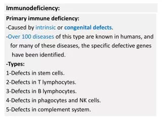
Immunodeficiency : Two types • Primary immunodeficiency: congenital or inherited • Secondary immunodeficiency: acquired • Regardless whether the cause is primary or secondary the spectrum of the disease will be the same
Humoral (B-cell) Immunodeficiencies • Caused by the improper production of one or all of the immunoglobins (antibodies) • Results in an increased of infections from Staphylococcus, Streptococcus, Haemophilus,and Pseudomonas. • Primary Humoral Immunodeficiencies include: • X-Linked (Bruton’s) Agammaglobinulinemia • Common Variable Immunodeficiency • Selective Immunoglobin A Deficiency • Secondary Humoral Immunodeficiencies: • General comments
Primary Humoral Immunodeficiency Disorders • X-Linked (Bruton’s) Agammaglobinulinemia: • Genetic disease that only affects males which results in undetectable levels of all serum immunoglobins • The genetic defect inhibits the differentiation of B-cells into plasma cells which ultimately are responsible for producing immunoglobins • Because of abnormally low immunoglobin levels, the child is susceptible to certain infections such as: • Meningitis • Recurrent ear infections • Chronic sinus/upper respiratory infections
Primary Humoral Immunodeficiency Disorders • Common Variable Immunodeficiency: • Similar to X-linked agammaglobulinemia • The terminal differentiation from mature B cells to plasma cells is blocked resulting in reduced serum immunoglobin levels • Symptoms are similar to agammaglobulinemia, but the onset occurs between 15 and 35 years. • Patients have a tendency for chronic lung disease, autoimmune disorders, hepatitis, gastric carcinoma, and chronic diarrhea associated with malabsorption.
Primary Humoral Immunodeficiency Disorders • Selective Immunoglobin A Deficiency: • Most common immunoglobin deficiency affecting 1 in 400 to 1 in 1000 persons • Main physical characteristic is a severe reduction in the levels of serum and secretory IgA that results from the inability of mature B cells to transform into IgA secreting plasma cells • Usually there are no overt S/S because IgG and IgM can compensate • Persons with this disease tend to be affected with: • Chronic upper respiratory infections • Chronic GI infections • Increased incidence of allergic diseases such as asthma
Secondary Humeral Immunodeficiency Disorders • Can occur as a consequence of selective loss of immunoglobulin through the gastrointestinal or genito-urinary tracts. • Example would be a person with Nephrotic syndrome who losses IgA and IgG in the urine because of abnormal glomerular filtration.
Cellular (T-cell) Immunodeficiencies • Caused by problem with T lymphocytes (both CD4+ helper cells and CD8+ cytotoxic killer cells) • Patient’s have more severe symptoms than with humeral immunodeficiencies • Children rarely survive beyond infancy or childhood • Primary Cell Mediated Immunodeficiency disorders include: • DiGeorge Syndrome • X-Linked Immunodeficiency with Hyper-IgM • Secondary Cell-Mediated immunodeficiency: • General comments
Primary Cell-Mediated Immunodeficiency Disorders • DiGeorge Syndrome: • Disease affecting both sexes • Caused by embryonic developmental defect resulting in the malformation of the: • Thymus gland • Parathyroid gland • Head/neck area • Heart • Associated facial abnormalities include increased distance between the eyes, upward bowing of upper lip (fish mouth), low-set/posteriorly angulated ears, split uvula • children are required to undergo blood transfusions/bone marrow transplants in order to survive
Primary Cell-Mediated Immunodeficiency Disorders • X-Linked Immunodeficiency with Hyper-IgM • Occurs only in males • Characterized by low levels of IgA and IgG and high levels of IgM • Once thought of a B cell disorder, however, now traced to T cells • There is a failure of T cells to signal B cells to produce IgA and IgG and instead only produce IgM • Children become symptomatic the first 2 years of life with recurrent pyogenic infections
Secondary Cell-Mediated Immunodeficiency • More common than primary cell-mediated immunodeficiency • Associated with acute viral infections (HIV, Herpesvirus) and with certain malignancies (Lymphoma, Hodgkin's disease).
Combined T-Cell and B-Cell Immunodeficiencies • Severe Combined Immunodeficiency (SCID) • Caused by diverse genetic mutations resulting in the absence of ALL immune function • Infants with this disease lead a short life (avg 2 years) with chronic opportunistic infections • There is a milder form known as combined immunodeficiency syndrome having a low, but not absent T-cell function. Antibody forming capacity is impaired, not absent. • Children are prone to recurrent pulmonary infections, failure to thrive, oral and cutaneous candidiasis, chronic diarrhea, recurrent skin infections, sepsis, and urinary tract infections. • Although they live longer than children with SCID they often die early in life.
Combined T-Cell and B-Cell Immunodeficiencies • Ataxia – Telangiectasia: • Results from a mutation on chromosome 11 • Condition consists of worsening ataxia (lack of coordination) and telangiectasia (dilated capillaries and arterioles) on the skin and conjunctiva. • Also results in endocrine and hepatic abnormalities also • Children have reduced levels of IgA, IgE, and IgG, and decreased ratio of CD4+ helper T cells to CD8+ cells. • Children are prone to recurrent upper and lower respiratory infections and an increased risk of malignancy. • Death from lymphoma is common
Combined T-Cell and B-Cell Immunodeficiencies • Wiskott-Aldrich Syndrome: • Patient has decreased IgM and elevated levels of IgA and IgE. • T-cell dysfunction is initially mild then progressively worsens making child susceptible to Hodgkin’s disease and lymphoma • They are also susceptible to infections (including septicemia and meningitis) caused by encapsulated microorganisms • Signs and symptoms: • eczema • chronic infections • low platelet counts • Bone marrow transplants have been helpful in these children
Disorders of Complement System • Complement system is an important part of the non-specific immune response. • Complement promotes chemotaxis, opsonization, and phagocytosis of invasive pathogens, bacteriolysis, and anaphylactic reactions. • Primary complement disorders are caused by genetic problems • Deficiency of C1 and C4 are not at increased risk for infection because alternate pathways can be activated. • These patients are more prone to autoimmune diseases such as Lupus Erythematosus. • Deficiency of C2: causes susceptibility to multiple life threatening bacteria
Disorders of Complement System • Secondary Disorders of Complement • Occur in persons with normal complement systems who have rapid activation or turnover of complement components • Can also occur with conditions where there is a decreased production complement components such as in liver cirrhosis or malnutrition
Disorders of Phagocytosis • Phagocytic system: • Composed primarily of: • Neutrophils • Eosinophils • Monocytes. • Phagocytic cells function to migrate to site of infection, adhere to the tissue, engulf invading material, and then digest it. • A defect in the phagocytic system results in a decreased number of phagocytic cells. • Persons are prone to bacterial infections, often by Candida and filamentous fungi.
Hypersensitivity Disorders • Hypersensitivity disorders are excessive or inappropriate activation of the immune system caused by immune responses to antigens that produce tissue-injury causing inflammation • hypersensitivity disorders are divided into 4 categories • Type 1Hypersensitivity reaction • Type 2 Hypersensitivity reaction • Type 3 Hypersensitivity reaction • Type 4 Hypersensitivity reaction
Type I Hypersensitivity (IgE-Mediated) Disorders • Type I reactions are immediate-type (within 30 min) hypersensitivity reactions that are triggered by the binding of an antigen to IgE located on the surface of a mast cell • Release of chemical mediators such as histamine produces an allergic reaction that stimulates the inflammatory process • Physiologic effects of histamine include: • Promotes vasodilation of blood vessels • Increases the permeability of blood vessels • Causes smooth muscle contraction • Causes bronchial constriction
Examples of Type I hypersensitivity disorders include: • Allergic Rhinitis: • Typical S/S include: • Sneezing • Itching • Watery discharge from eyes/nose • Typical antigens include: • Pollens from ragweeds, grass, weeds • Fungal spores • House dust mites • Animal dander/feathers • Food Allergies: the most common food allergies include: milk, peanuts, shellfish, and eggs • Food allergies may produce a severe allergic response called anaphylaxis
Type II-Antibody Mediated Disorders: • AKA cytotoxic responses • Result from the interaction between IgG and IgM antibodies and antigens which results in the activation of cytotoxic immune cells • Examples include: • Mismatched blood transfusions • Hemolytic disease
Type III- Immune Complex Disorders: • Caused by the formation of insoluble antigen/antibody complexes that adversely increases immune function and result in the release of certain chemicals that cause tissue damage • Most common areas of the body affected by Type II disorders include: • Renal glomerulus • Lung • Synovial membrane of joints • The two most common examples of Type II disorders include: • Rheumatoid arthritis (RA) • Systemic Lupus Erythematosis (SLE)
Type IV- Cell Mediated Disorders: • Delayed type of reaction that is mediated by T-cells not antibodies • Usually occur 24 to 72 hours after antigen exposure • Mediated by T lymphocytes • Examples include: • Allergic Contact Dermatitis- is an inflammatory response confined to the skin that results when the skin comes in contact with an irritating substance • usually produces inflamed blisters on skin • common causes include exposure to: 1. poison ivy 2. cosmetics 3. topical medications 4. latex
Autoimmune Disease • Def: Self-tolerance: to function properly the immune system must be able to distinguish foreign antigens and self-antigens • Autoimmune disease- occurs when the immune system cannot differentiate antigens from self-antigens and begins to attack host tissue of the body that results in localized or systemic injury • Examples of autoimmune diseases include: • Myasthenia gravis (MG) • Rheumatoid arthritis (RA) • Systemic Lupus Erythematosis (SLE ) • Graves’ Disease
Loss of B-Cell Tolerance: results in autoimmune diseases that produce autoantibodies: • Example is Graves disease: produces hyperthyroidism due to autoantibodies that stimulate the TSH receptors on the thyroid gland. • Loss of T-Cell Tolerance: results from the release of self-reactive T cells from the thymus. • Normally T-cells that have a high affinity for host cells undergo apoptosis. If these cells do not die they leave the thymus gland and cause tissue damage.
HIV AIDS • The pathogen of AIDS is HIV (Human Immunodeficiency Virus) • At the end of 2006: • Nearly 40 million worldwide were living with HIV/AIDS • 25 million people had died of the infection • 4.3 million new infections (2006) • 50% of these women and children • Projected: over the 10 next years there will be 100 million people infected with HIV. • Most new infections: people under 25 in developing countries. • Sub-Sarahan Africa will be most affected.
The AIDS Epidemic • S/S associated with decreased immune function were first recognized in homosexual men in 1981 • Although initially thought to affect the gay male population specifically, AIDS cases began to be diagnosed in the following high risk groups: • IV drug users • Hemophiliacs • Blood transfusion recipients • High risk heterosexuals • As a result of HIV infection, the body’s immune system begins to progressively fail making the infected person more susceptible to infections that are normally harmless to the body (Opportunistic infections)
Transmission of HIV • Sexual contact: • Most frequent mechanism • 75-80% via unprotected sex • The virus can pass from semen and vaginal secretions into the bloodstream through mucous membranes and wounds in the skin. • Blood-to-blood contact: • Needles, syringes, or Transfusions • Perinatally: • Transmission can occur in utero, during labor and delivery, or by breast feeding • Studies still show that HIV is not spread through casual contact nor by vectors such as mosquitoes
Def: Seroconversion: The point where an infected person converts from being negative for HIV antibodies to having HIV antibodies • Occurs 1 to 3 months after exposure to HIV. • But can take up to 6 months • Def: Window period: The period of time after infection but before seroconversion. • During this time the patient does not have symptoms, nor antibodies for HIV that can be picked up on a blood test. • This patient can unknowingly transmit the disease • As a result the FDA requires collection center to screen potential donors through interviews designed to identify behavior known to present risk for HIV infection.
Pathophysiology and Clinical Course • 2 forms of HIV • HIV 1 • Most associated with AIDS in the US, Europe, and Central Africa • HIV 2 • Prevalent in West Africa • Spreads more slowly in comparison to HIV 1 • Causes disease more slowly in comparison to HIV 1 • Cells attacked by HIV • CD4+ T lymphocytes • Macrophages • Dendritic cells: specialized protective cells found in tissues where antigens enter the body
The HIV Virus • HIV is a retrovirus: carries its genetic information on RNA • The virus is spherical in shape with a core in the center surrounded by a lipid envelope (membrane) • Core: contains • A protein (p24) • Two copies of RNA • 3 viral enzymes: • Protease • Reverse transcriptase • Integrase • Envelope: contains a protein matrix called p17 which lies directly beneath the viral envelope. Viral envelope is studded by 2 viral glycoproteins, gp120 and gp41 which are important for the infection process
Life cycle of the HIV-1 Eight steps 1. Attachment of the HIV virus to CD4+ receptor: a. Virus attaches to the surface of a CD4+ cell b. The virus then binds its gp 120 and gp 41 glycoproteins with chemokine receptors on the CD4+ cell 2. Internalization: a. Once the virus has attached, viral peptides fuse with the CD4+ cell membrane causing an uncoating of the virus. b. Viral contents (viral RNA, reverse transcriptase, integrase, and protease) then enter the cell.
3. Reverse transcription: a. Viral RNA must be converted to DNA (in order to reproduce) b. Done by the enzyme reverse transcriptase which produces a mirror image of the viral RNA c. The result is a double-stranded DNA molecule 4. Integration: a. Newly created viral DNA enters nucleus of CD4+ b. Viral DNA becomes part of the cell DNA using integrase enzyme 5. Transcription of inserted viral DNA: a. Inserted viral DNA to produce viral mRNA 1. mRNA has the instructions for building new HIV viruses
6. Translation of viral messenger: a. rRNA uses the instructions of mRNA to create viral proteins and enzymes known as polyprotein 1. Polyprotein enzymes are needed to construct viral components into new viruses 7. Cleavage: a. During this stage protease enzymes cut viral proteins into single proteins b. These proteins will make up the new viruses
Assembly and release: • a. Proteins and viral RNA are assembled into new viruses • b. New viruses are built inside the cell resulting in the death of • the CD4+ cell • c. When the CD4+ cell dies, the viral particles (virions) are • released into the bloodstream to infect other CD4+ cells. • d. Every day millions of CD4+ cells are destroyed, releasing • billions of viral particles into the blood • e. The immune system is able to keep up with the infection by • producing new T-cells, however, over years, the T-cell count • decreases and the amount of virus in the blood increases. • f. Until a person’s T-cell count falls to a very low level, a • person can remain asymptomatic, even though viral • replication is taking place.
Infected helper T-cell; the small blue globules are HIV particles.
Classification of HIV • 3 Categories relating to the amount of CD4+ cells per microliter of blood (normal is 800 to 1000 cells/microliter) • Category 1: >500 cells/microliter • Category 2: 200 to 499 cells/mircroliter • Category 3: <200 cells/microliter. • 3 Clinical categories based on symptoms: • Clinical Category A: persons who are asymptomatic or have persistent generalized lymphadenopathy, or symptoms of primary infection • Clinical Category B: person has symptoms of immune deficiency, however, not severe enough to be AIDS • Clinical Category C: person has AIDS defining illnesses such as pneumocystis carinii pneumonia.
Phases of HIV • 3 phases which occur over 8 to 12 years • Primary Infection phase: • Signs and symptoms appear 2 to 4 weeks after exposure lasting a few days to 2 weeks • Patient has fever, fatigue, myalgia, sore throat, night seats, GI problems, lymphadenopathy, maculopapular rash, and headache. • During this phase there is a high amount of viral replication (up to 1 million/ml) and a decreasing amount of CD4+ • After a few weeks the immune system catches up controlling viral replication where it remains for several years. • If diagnosed in this stage and therapy is started treatment may reduce the number of long-living CD4+ infected cells
Chronic Asymptomatic (Latency Phase) • Patient has no signs or symptoms of illness • On average this phase lasts 10 years • There is a decreasing amount of CD4+ cells down to 200 per microliter or lower. • AIDS Phase • Occurs when the CD4+ cell count falls less than 200/microliter • Patient is susceptible to opportunistic infections • Without antiretroviral therapy this phase can lead to death within 2 to 3 years
Clinical Course of HIV • Typical progressors: 60% to 70% acquire AIDS 10 to 11 years after infection • Rapid progressors: 10% to 20% progress rapidly and acquire AIDS in less than 5 years • Slow progressors: 5% to 15% are slow progressors, who do not progress to AIDS for more than 15 years • Long-term non-progressors: 1% have been infected for at least 8 years, are antiretroviral naive, have high CD4+ cell counts and usually very low viral loads
Opportunistic Infections Affecting AIDS • Bacterial opportunistic infections • Bacterial pneumonia, TB , salmonella, mycobacterium avium-intracellulare complex (MAC) • Fungal opportunistic infections • Candidiasis, coccidiomycosis, cryptococcosis, and histoplasmosis • Protozoal opportunistic infections • Cryptosporidiosis, isosporiasis, pneumocystiasis, and toxoplasmosis • Viral infections • Cytomegalovirus (CMV), herpes, and progressive multifocal leukoencephalopathy (PML)
Most Common Opportunistic Infections Affecting AIDS in the U.S. • Pneumocystis carinii pneumonia (PCP) • CMV • Oropharyngeal or esophageal candidiasis (thrush) • Infections caused by MAC (mycobcacterium avium-intrcellulare complex)
Clinical Manifestations 1. Respiratory Manifestations: the most common respiratory diseases in person with AIDS include: a) Pneumocystis carinii pneumonia (PCP)- is caused by a protozoa that is common in soil, and houses • patients tend to complain of: • Cough, fever, shortness of breath, and weight loss b) Mycobacterium tuberculosis: is the leading cause of death for people with AIDS worldwide • infections site is primarily in the lungs but may also be found in the kidneys and bone marrow • common S/S include: fever, night sweats, cough and weight loss
2) Gastrointestinal Manifestations- diseases of the GI tract are the most frequent complications of AIDS and usually include: a) Esophageal Candidiasis (Thrush)- presents as cheesy matter along the entire GI tract including the mouth b) Diarrhea (Gastroenteritis)- is the most common complaint in persons with AIDS and is usually caused by the protozoa Cryptosporidium parvum
3) Nervous System Manifestations: due to severe immune compromise, both the CNS and PNS are susceptible to several neurological disorders that can include: a) AIDS Dementia Complex- is a syndrome involving both cognitive and motor system dysfunction that is caused by the pathological effects of HIV itself • typical S/S include: • loss of attention span/concentration • decrease in mental speed and agility • decrease in overall motor function • apathetic behavior b) Toxoplasmosis- is an opportunistic parasitic infection that affects the CNS • typical S/S include: • fever • headache • confusion/lethargy • visual disturbances • seizure activity