Download
1 / 92
990 likes | 1.74k Vues
Bacillus anthracis. Hirotaka Ishibashi Jennifer Jolivet Sean Patrick Kelly. Bacillus anthracis. Gram + rod Facultative anaerobe 1 - 1.2 µ m in width x 3 - 5 µ m in length Belongs to the B. cereus family Thiamin growth requirement Glutamyl-polypeptide capsule Nonmotile

E N D
Bacillus anthracis Hirotaka Ishibashi Jennifer Jolivet Sean Patrick Kelly
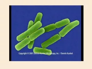
Bacillus anthracis • Gram + rod • Facultative anaerobe • 1 - 1.2µm in width x 3 - 5µm in length • Belongs to the B. cereus family • Thiamin growth requirement • Glutamyl-polypeptide capsule • Nonmotile • Forms oval, centrally located endospores http://www.bact.wisc.edu/Bact330/lectureanthrax
Endospore • Oxygen required for sporulation • 1 spore per cell • dehydrated cells • Highly resistant to heat, cold, chemical disinfectants, dry periods • Protoplast carries the material for future vegetative cell • Cortex provides heat and radiation resistance • Spore wall provides protection from chemicals & enzymes http://www.gsbs.utmb.edu/microbook/ch015.htm
Genetics • 1 chromosome • 5.2 million bp • Ames strain sequenced • 2 plasmids • px01 • 184 kbp • Pathogenicity island • pX02 • 95.3 kbp • Capsule • Anthrax receptor • Occurs > than ten thousendfold on macrophage cell • ATR/TEM8 gene • Chromosome 4 gib.genes.nig.ac.jp/single/ main.php?spid=Bant_AME
Where is Anthrax? http://www.vetmed.lsu.edu/whocc/mp_world.htm
Anthrax • From the Greek word anthrakos for coal • Caused by spores • Primarily a disease of domesticated & wild animals • Herbivores such as sheep, cows, horses, goats • Natural reservoir is soil • Does not depend on an animal reservoir making it hard to eradicate • Cannot be regularly cultivated from soils where there is an absence of endemic anthrax • Occurs sporadically throughout US • South Dakota, Arkansas, Texas, Louisiana, Mississippi, California recognized endemic areas • Anthrax zones • Soil rich in organic matter (pH < 6.0) • Dramatic changes in climate
Anthrax Infection & Spread • May be spread by streams, insects, wild animals, birds, contaminated wastes • Animals infected by soilborne spores in food & water or bites from certain insects • Humans can be infected when in contact with flesh, bones, hides, hair, & excrement • nonindustrial or industrial • cutaneous & inhalational most common • Risk of natural infection 1/100,000 • Outbreaks occur in endemic areas after outbreaks in livestock
Three forms of Anthrax • Cutaneous anthrax • Skin • Most common • Spores enter to skin through small lesions • Inhalationanthrax • Spores are inhaled • Gastrointestinal (GI)anthrax • Spores are ingested • Oral-pharyngeal and abdominal
Milestones in Anthrax History • Early history • 1800s • 1900s • Recent years • Outbreaks in Thailand and US
History of Anthrax (Early history) • Although anthrax dates back more than 3,000 years, it was not recognized as a disease until the 18th century. • 1500 B.C - A “plague of boils” in Egypt affected the Pharaoh’s cattle. ‘Boils’ are symptomatic of anthrax. • 1600s - The “Black Bane” thought to be anthrax, in Europe kills over 60,000 cattle. • 1700s - There are some accounts of human cases.
History (1800s) • Early 1800s - The first human cases of cutaneous anthrax in the US and UK were reported in men who contracted the disease after having been in contact with infected livestock. • The disease was called Wool Sorter’s disease or Rag Picker’s disease because it affected workers in those trades. • 1868 - Anthrax was observed under a microscope. • 1876 - German bacteriologist Robert Koch confirmed bacterial origin of anthrax.
History (Early 1900s) • 1915 - German agents injected horses, mules, and cattle with anthrax during WWI. This was the first recorded use of anthrax as a biologicalweapon. • 1937 - Japan started a biological warfare program in Manchuria, including tests involving anthrax. • 1942 - UK demonstrated experiments using anthrax at Gruinard Island off the coast of Scotland. • 1943 - United States began developing anthrax weapons. • 1945 - In Iran an anthrax outbreak killed more than 1 million sheep.
History (Late 1900s) • 1950s and 60s - U.S. biological warfare program continues after WWII at Fort Detrick, Maryland • 1969 - President Nixon ended United States' offensive biological weapons program, but defensive work still continues. • 1970 - Anthrax vaccine for humans was approved by U.S. FDA. • 1978-80 - The world's largest outbreak of human anthrax via insect vectors or contaminated meat struck Zimbabwe, Africa where more than 10,000 cases were recorded and over 180 people died. • 1979 - In Soviet Union, aerosolized anthrax spores were released accidentally at a military facility, affecting 94 and killing 64 people.
History (Recent years) • 1991 - About 150,000 U.S. troops were vaccinated for anthrax in preparation for Gulf War. • 1990-93 - The cult group, Aum Shinrikyo, released anthrax spores in Tokyo, fortunately no one was injured. On February 27, 2004, the leader of this group was given a sentence of death at a district court in Tokyo. • 1995 - Iraq produced 8,500 liters of concentrated anthrax as part of the biological weapon program under Saddam Hussein’s administration. • 2001 - Letters containing anthrax spores were mailed to many places in the US such as NBC, New York Times, and Media in Miami. In Florida, a man died after inhaling anthrax at the office.
Outbreaks in Thailand • This picture is 9 days after the onset of symptoms of oral-pharyngeal anthrax. • 1982 - In rural Northern Thailand, an outbreak of 52 cases of cutaneous anthrax and 24 cases of oral-pharyngeal anthrax occurred. • Oral-pharyngeal anthrax: an unusual manifestation of humaninfection with B. anthracis. • 1987 - 14 cases of both oral- pharyngeal and abdominal anthrax occurred. • Caused by the consumption of contaminated water and buffalo meat. Thira Sirisanthana, Arthuer Brown, Anthrax of the Gastrointestinal Tract, Emerging Infectious Diseases, Vol. 8, 7, July 2002
Outbreaks in the US • In the early 1900’s approximately 130 cases occurred annually due to the following reasons. 1) Agricultural, farm workers exposed to infected animals 2) Processors exposed to infected animal products (hair, leather, wool, bone) 3) Laboratory workers contacted with anthrax spores 4) Civilians exposed to contaminated imported animal products • These four are rare today.
in 1957 animal vaccination started Natural Outbreaks in the U.S. (1951–2003) in 1970 human vaccination started 2003 • N = 409 • 391 cases were cutaneous anthrax. • 18 cases were inhalation anthrax • GI anthrax has not been recognized yet • Since 1990 only 2 cases of cutaneous anthrax of naturally • occurring infection have been reported. Anthrax Overview PPT, CDC, 2001 (http://www.cdc.gov/)
Natural Outbreaks in North Dakota • The highest occurrence of Anthrax outbreaks in the US • 1989-1999 - 26 cases of infected livestock were reported. • 2000 - 33 cases were reported during July-September. • Total of 180 animals (beef cattle, horses, and bison) died and one person was infected with cutaneous anthrax. • Roughly, cases appear every 2 years in North Dakota
Pathogenesis • The infectious dose of B. anthracis in humans by any route is not precisely known. • Rely on primate data • Minimum infection dose of ~ 1,000-8,000 spores • LD50 of 8,000-10,000 spores for inhalation • Virulence depends on 2 factors • Capsule • 3 toxins http://www.kvarkadabra.net/index.html?/biologija/teksti/biolosko_orozje.htm
Capsule • Glycocalyx • Sticky, gelatinous polymer external to cell wall • pX02 plasmid • Made up of D-glutamic acid • Non-toxic on its own • Only encapsulated B. anthracis virulent • Most important role during establishment of disease • Protects against phagocytosis & lysis during vegetative state http://textbookofbacteriology.net/BSRP.html
Toxins • pX01 plasmid • AB model • Binding • Activating • Protective antigen (PA), edema factor (EF) & lethal factor (LF) • Make up 50% of proteins in the organism • Individually non-toxic • PA+LF lethal activity • EF+PA edema • EF+LF inactive • PA+LF+EF edema & necrosis; lethal http://www.rcsb.org/pdb/molecules/pdb28_1.html
Toxins (2) • Protective antigen (PA, 83kDa) • Pag gene • Binds to receptor & helps internalize other 2 proteins • Edema factor (EF, 89 kDa) • Cya gene • Adenylate cyclase • Affects all cells • Lethal factor (LF, 87 kDa) • Lef gene • More important virulence factor • Metalloprotease • Cleaves mitogen activated protein kinase kinsase (MAPKK) • Affects only macrophages http://www.ericse.org/anthrax/anthraxmicrographs.html
Mechanism of Infection • Anthrax spores enter body • Germinate & multiple in lymph nodes • PA, EF, LF excreted from bacteria • PA binds to TEM8. • PA nicked by protease furin • 20-kDa segment off leaving 63-kDa peptide • Heptamer forms • EF and/or LF binds • Complex internalized by endocytosis • Acidification of endosome • LF or EF crosses into cytosol via PA mediated ion-conductive channels • LF cleaves MAPKK 1 & 2 • EF stimulates cAMP http://kugi.kribb.re.kr/KUGI/Pathways/BioCarta/anthraxPathway/
Outcome • Do not understand exactly how symptoms occur • EF converts ATP to cAMP • Increases cAMP levels over 1,000 fold • Impairs neutrophil function • Alters water homeostasis • Edema • LF cleaves MAPKK at its N terminus • Disrupts pathways involved in cell growth & maturation • Increased synthesis of tumor necrosis factor-α & interleukin-1β • Macrophage lysis • More cells infected with bacteria & toxin • Septic shock & death • Death probably results from high levels of bacteria secreting LF toxins in blood • At death, blood contains as many as 109 bacilli/ml (depending on the species)
Regulators • Bicarbonate or CO2 stimulates capsule and PA formation • LF requires zinc ions • EF requires calmodulin, a major intracellular calcium receptor • Transcriptional regulator AcpA on pX02 controls expression of capsule • atxA on pX01 is a positive regulator necessary for transcription of all 3 toxin genes
Clinical Information • Infection • Symptoms (1st and 2nd phase) • Three forms of Anthrax infection and their Pathology • Diagnosis
Infection of Anthrax • The estimated number of naturally occurring human cases of anthrax in the world is 20,000 to 100,000 per year. • Humans are infected through contact with infected animals and their products because of human intervention. • Anthrax spores contaminate the ground when an affected animal dies and can live in the soil for many years. • Anthrax can also be spread by eating undercooked meat from infected animals. • Anthrax is NOT transmitted from person to person. • Humans can be exposed but not be infected.
What are the symptoms for anthrax? • There are two phases of symptom. • 1) Early phase - Many symptoms can occur within 7 days of infection • 2) 2nd phase - Will hit hard, and usually occurs within 2 or 3 days after the early phase.
- Early Phase Symptoms - • Fever (temperature > 100 degrees F) • Chills or night sweats • Headache, cough, chest discomfort, sore throat • Joint stiffness, joint pain, muscle aches • Shortness of breath • Enlarged lymph nodes, nausea, loss of appetite, abdominal distress, vomiting, diarrhea • Meningitis
- 2nd Phase Symptoms - • Breathing problems, pneumonia • Shock • Swollen lymph glands • Profuse sweating • Cyanosis (skin turns blue) • Death
Three clinical forms of Anthrax • 3 types of anthrax infection occur in humans: 1) Cutaneous 2) Inhalation 3) GI
Cutaneous Anthrax • 95% of anthrax infections occur when the bacterium enters a cut or scratch on the skin due to handling of contaminated animal products or infected animals. • May also be spread by biting insects that have fed on infected hosts. • After the spore germinates in skin tissues, toxin production initially results in itchy bump that develops into a vesicle and then painless black ulcer. http://science.howstuffworks.com/anthrax1.htm
Cutaneous Anthrax (2) • The most common naturally occurring form of anthrax. • Ulcers are usually 1-3 cm in diameter. • Incubation period: • Usually an immediate response up to 1 day • Case fatality after 2 days of infection: • Untreated (20%) • With antimicrobial therapy (1%)
Cutaneous Anthrax (3) CDC, Cutaneous Anthrax—Vesicle Development
Inhalation Anthrax • The infection begins with the inhalation of the anthrax spore. • Spores need to be less than 5 microns (millionths of a meter) to reach the alveolus. • Macrophages lyse and destroy some of the spores. • Survived spores are transported to lymph nodes. • At least 2,500 spores have to be inhaled to cause an infection. Inhalation Anthrax, Introduction, DRP, Armed Forces Institute of Pathology
Inhalation Anthrax (2) • Disease immediately follows germination. • Spores replicate in the lymph nodes. • The two lungs are separated by a structure called the mediastinum, which contains the heart, trachea, esophagus, and blood vessels. • Bacterial toxins released during replication result in mediastinal widening and pleural effusions (accumulation of fluid in the pleural space). Inhalation Anthrax, Introduction, DRP, Armed Forces Institute of Pathology
Inhalation Anthrax (3) • Death usually results 2-3 days after the onset of symptoms. • Natural infection is extremely rare (in the US, 20 cases were reported in last century). • Inhalation Anthrax is the most lethal type of Anthrax. • Incubation period: • 1–7 days • Possibly ranging up to 42 days (depending on how many spores were inhaled). • Case fatality after 2 days of infection: • Untreated (97%) • With antimicrobial therapy (75%)
Gastrointestinal Anthrax • GI anthrax may follow after the consumption of contaminated, poorly cooked meat. • There are 2 different forms of GI anthrax: 1) Oral-pharyngeal 2) Abdominal • Abdominal anthrax is more common than the oral-pharyngeal form. http://science.howstuffworks.com/anthrax1.htm
GI Anthrax (2) • Oral-pharyngeal form - results from the deposition and germination of spores in the upper gastrointestinal tract. • Local lumphadenopathy (an infection of the lymph glands and lymph channels), edema, sepsis develop after an oral or esophageal ulcer. • Abdominal form - develops from the deposition and germination of spores in the lower gastrointestinal tract, which results in a primary intestinal lesion. • Symptoms such as abdominal pain and vomiting appear within a few days after ingestion.
GI Infection (3) • GI anthrax cases are uncommon. • There have been reported outbreaks in Zimbabwe, Africa and northern Thailand in the world. • GI anthrax has not been reported in the US. • Incubation period: • 1-7 days • Case fatality at 2 days of infection: • Untreated (25-60%) • With antimicrobial therapy (undefined) due to the rarity
How is anthrax diagnosed? • Gram stain • Culture of B. anthracis from the blood, skin lesions, vesicular fluid, or respiratory secretions • X-ray and Computed Tomography (CT) scan • Rapid detection methods - PCR for detection of nucleic acid - ELISA assay for antigen detection - Other immunohistochemical and immunoflourescence examinations - These are available only at certain labs
Gram Stain Analysis • Useful for cutaneous and inhalation anthrax. • A blood sample or skin lesion is taken from the patient and cultured for 6 to 24 hours. • Gram stain takes about 10 to 15 minutes. • Identify whether the bacteria come from the anthrax category. Bacillus anthracis in Gram stain
Chest X-ray • Useful for inhalation and GI anthrax • Chest X-rays is advised as an initial method of inhalation anthrax detection, but it is sometimes not useful for patients without symptoms. • Find a widened mediastinum and pleural effusion. • Picture shows widened mediastinum caused by B. anthracis infection, resulting less available space in lungs At day 1 At day 3 Inhalation Anthrax, Introduction, DRP, Armed Forces Institute of Pathology
CT scan • Useful for inhalation and GI anthrax • Even when X-rays are negative, CT scans may provide more precise information. • Chest CT (Right) shows the increase in the size of the pleural effusions (accumulation of fluid in the pleural space). Inhalation Anthrax, Introduction, DRP, Armed Forces Institute of Pathology
PCR Assay • PCR is a target amplification method of nucleic acid based B. anthracis detection. • Used for the detection of anthrax toxin genes. ex) rpoB gene - used as a specific chromosomal marker for RT-PCR detection. • The rpoB gene was sequenced from 36 Bacillus strains • The assay was specific for 144 Bacillus anthracis strains from different geographical locations. • Provided 100% sensitivity and specificity
PCR Assay (2) • Detection time: - PCR only takes several hours ex) Rapid-cycle RT-PCR can be finished within 1-2 hours • Can start early treatment of Anthrax • There are many different types of PCR assays for the detection of Anthrax such as multiplex PCR, enterobacterial repetitive intergenic consensus-PCR (ERIC-PCR), and long-range repetitive element polymorphism-PCR. • Rapid diagnostic methods provide answers in minutes or hours instead of days.
Distinguishing inhalation Anthrax from cold or influenza • Anthrax, cold, and influenza patients have similar symptoms at early phase such as flu-like symptoms (fever, chills, cough, and muscle aches etc.) • Symptoms of Anthrax do not include a runny nose, which is common in cold and influenza . • Anthrax involves severe breathing problems and more vomiting. These symptoms are not very common in cold or influenza. • Anthrax have high white blood cell counts and no increase in the number of lymphocytes. • Flu usually have low white blood cell counts and an increase in the number of lymphocytes. • Inhalation anthrax has abnormality in X-ray or CT scan
Treatment • Before 2001, 1st line of treatment was penicillin G • Stopped for fear of genetically engineered resistant strains • 60 day course of antibiotics • Ciprofloxacin • fluoroquinolone • 500 mg tablet every 12h or 400 mg IV every 12h • Inhibits DNA synthesis • Doxycycline • 6-deoxy-tetracycline • 100 mg tablet every 12h or 100 mg IV every 12h • Inhibits protein synthesis • For inhalational, need another antimicrobial agent • clindamycin • rifampin • chloramphenico http://nmhm.washingtondc.museum/news/anthrax.html
Vaccine • BioThrax/Anthrax vaccine absorbed • Made by Bioport • Route of exposure not important • Administered subcutaneously • .5mL at 0, 2, and 4 weeks, and at 6, 12, & 18 months, & booster doses at 1 yr intervals • PA from attenuated, nonencapsulated Sterne strain absorbed onto aluminum hydroxide • Contains no dead or live bacteria in the preparation • Antibodies to PA prevent binding to the target cell & confer protection from anthrax. • 95% of vaccinated Rhesus monkeys survived lethal doses of inhaled anthrax • A December 22, 2003 ruling temporarily halted the Department of Defense’s anthrax vaccination program • Lifting of that injunction on January 7, 2004
Who gets it? • People who work directly with it in the lab • People who work with imported animal hides or furs in areas where standards are insufficient to prevent exposure to anthrax spores. • People who handle potentially infected animal products in high-incidence areas • Military personnel deployed to areas with high risk for exposure to the organism.