Download
1 / 40
400 likes | 656 Vues
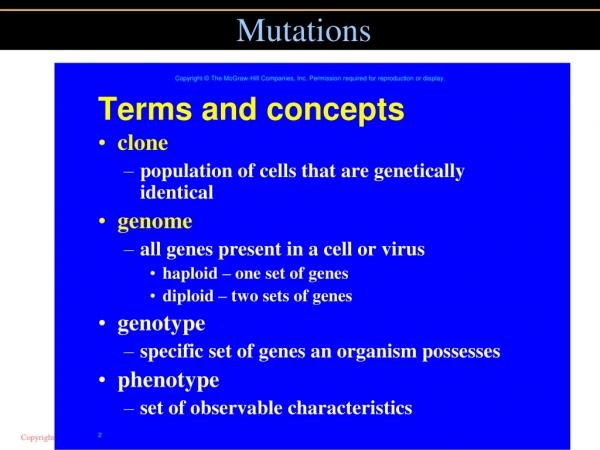
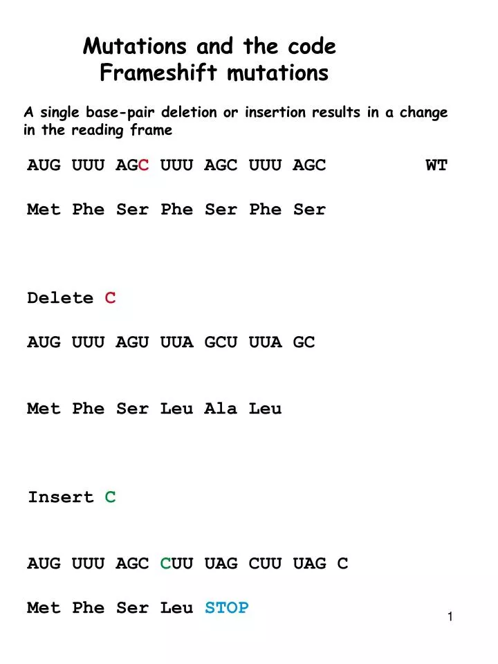
Mutations and the code Frameshift mutations. A single base-pair deletion or insertion results in a change in the reading frame. AUG UUU AG C UUU AGC UUU AGC WT Met Phe Ser Phe Ser Phe Ser Delete C AUG UUU AGU UUA GCU UUA GC Met Phe Ser Leu Ala Leu Insert C

E N D
Mutations and the code Frameshift mutations A single base-pair deletion or insertion results in a change in the reading frame AUG UUU AGC UUU AGC UUU AGC WT Met Phe Ser Phe Ser Phe Ser Delete C AUG UUU AGU UUA GCU UUA GC Met Phe Ser Leu Ala Leu Insert C AUG UUU AGC CUU UAG CUU UAG C Met Phe Ser Leu STOP
Frameshift mutations- Deletion A single base-pair deletion or insertion results in a change in the reading frame AUG UUU AGC UUU AGC UUU AGC Met Phe Ser Phe Ser Phe Ser Delete C Delete GC Delete AGC
Frameshift mutations-Insertion A single base-pair deletion or insertion results in a change in the reading frame AUG UUU AGC UUU AGC UUU AGC Met Phe Ser Phe Ser Phe Ser Insert C Insert CC Insert CCC
Missense mutations Missense mutations alters ONE codon so that it encodes a different amino acid UUU UUU UGC UUU UUU WT UUU UUU UGG UUU UUU mut
Consequences of Missense Mutations Missense mutations alter one of the many amino acids that make a protein Its consequences depend on which amino acid is altered Conservative mutations: K to R Nonconservative mutations: K to E Surface Vs buried Mutations in globular domains Vs un structured tails Silent mutations Mutations in non-coding regions Nonsense mutations
Silent Mutations Silent mutations do not alter the amino acid sequence! AUG UUU AGC UUU AGC UUU AGC WT AUG UUC AGC UUU AGC UUU AGC Mut Mutations that occur in introns are also silent Mutations that occur in non-genic regions are silent
Mutations in non-protein coding regions Mutations in the promoter or ribosome binding site are also mutagenic Reduced expression of mRNA might result in reduced levels of proteins Mutations in splicing junctions may also be mutagenic Improperly spliced mRNA will result in the intron being Translated Mutations in tRNA or aminoacyl-tRNA synthase are mutagenic
Nonsense mutations Nonsense mutations alter one codon so that it now encodes for a STOP codon UUU UUU UGC UUU UUU Phe Phe Cys Phe Phe UUU UUU UGA UUU UUU Phe Phe STOP Nonsense mutations insert a stop codon which results in premature termination Truncated polypeptide usually results in loss of function for polypeptide
Nonsense suppressor mutations! These are the result of a mutation in the anti-codon loop of a specific tRNA It allows the tRNA to recognize a nonsense codon and base pair with it. DNA Gene encoding tRNATRP Point mutation occurs in the anticodon loop This allows this tRNA to base pair with a stop codon and ?
Met Ala Phe Phe Trp AUC AAA Nonsense suppressor --- UUU UUU UAG UUU UUU ----- --- Phe Phe STOP Trp-tRNA has mutation In anticodon This allows it to pair with a stop codon 5’--- UUU UUU UAG UUU UUU -----3’ --- Phe Phe Trp Phe Phe ----> A mutant protein that is larger than normal will be synthesized!!
Met Ala Phe Phe Phe Phe Trp Trp AUC AUC AAA AAA AAA ---UAG--- 5’--- UUU UUU UAG UUU UUU -----3’ Nonsense and Nonsense suppressor --- UUU UUU CAG UUU UUU ----- --- Phe Phe Gln Phe Phe --- Nonsense mutation --- UUU UUU UAG UUU UUU ----- --- Phe Phe STOP What will happen if an individual carries both a nonsense mutation in a gene and a nonsense suppressor mutation in the anticodon loop of one of the trp-tRNA genes.
Spontaneous mutations Replication induced mutations of DNA Usually base substitutions Most spontaneous errors are corrected Mutations during meiotic pairing Small additions and deletions Environment induced changes Exposure to physical mutagens - radioactivity or chemicals Depurination (removal of A or G) Repair results in random substitution during replication Deamination (removal of amino group of base) (nitrous acid) Cytosine--uracil--bp adenine--replication-- Oxidation (oxoG) guanine--oxoguanine--bp adenine--replication -- Base analog incorporation during replication BU-T Intercalating agents X-rays- Generation of mutations
Methods used to study mutations Gross chromosomal changes- deletions, insertions, inversions, translocations Cytology- microscopy- karyotype Point mutations Small deletions, insertions Recombinant DNA technologies
Recombinant DNA technology When genes are mutated - proteins are mutated- DISEASE STATES OCCUR Sickle cell Anemia Globin 2 alpha globin chains 2 beta globin chains Mol wt 16100 daltons xfour = 64650 daltons Single point mutation in beta-globin Converts Glu to Val at position6 Need to know mutation Need to look at genes of individuals Genes lie buried in 6billion base pairs of DNA (46 chromosomes). Molecular analyses necessary Take advantage of enzymes and reactions that naturally occur in bacteria
Why all the Hoopla? Why all the excitement over recombinant DNA? It provides a set of techniques that allows us to study biological processes at the level of individual proteins in individuals! It plays an essential role in understanding the genetic basis of cancer in humans Recently found that mutations in a single gene called p53 are the most common Genetic lesion in cancers. More than 50% of cancers contain a mutation in p53 Cells with mutant p53 Chromosomes fragment Abnormal number of chromosomes Abnormal cell proliferation!
p53 To understand the complete biological role of p53 protein and its mutant phenotype we need to study the gene at multiple levels: Genetics- mutant gene- mutant phenotype Now what? Genetics will relate specific mutation to specific phenotype It usually provides No Information about how the protein generates the phenotype For p53 We would like to know The nucleotide sequence of the gene and the mutation that leads to cancer When and in which cells the gene is normally expressed (in which cells is it transcribed) At the protein level--Amino acid sequence Three-dimensional structure Interactions with other proteins Cellular information Is the location in the cell affected How does it influence the behavior of the cell during division Organism phenotype
Alkaptonuria Degenerative disease. Darkening of connective tissue, arthritis Darkening of urine Garrod characterized the disorder- using Mendels rules- Autosomal recessive. Affected individuals had normal parents and normal offspring. 1908 Garrod termed the defect- inborn error of metabolism Homogentisic acid is secreted in urine of these patients. This is an aromatic compound and so Garrod suggested that it was an intermediate that was accumulating in mutant individuals and was caused by lack of enzyme that splits aromatic rings of amino Acids. 1958 La Du showed that accumulation of homogentistic acid is due to absence of enzyme in liver extracts 1994 Seidman mapped gene to chromosome 3 in human 1996 Gene cloned and mutant identified P230S &V300G 2000 Enzyme principally expressed in liver and kidneys
Basic techniques --- Nucleic acid hybridization complementary strands will associate and form double stranded molecules --- Restriction Enzymes These enzymes recognize and cleave DNA at specific sequences --- Blotting Allows analysis of a single sequence in a mixture --- DNA cloning This allows the isolation and generation of a large number of copies of a given DNA sequence --- DNA sequencing Determining the array of nucleotides in a DNA molecule --- PCR --- Transformation Stably integrating a piece of DNA into the genome of an organism --- Genetic engineering Altering the DNA sequence of a given piece of DNA --- Genomics Analyzing changes in an entire genome
Nucleic acid hybridization Complementary strands of DNA or RNA will specifically associate DNA is heated to 100C, the hydrogen bonds linking the two strands are broken The double helix dissociates into single strands. As the solution is allowed to cool, strands with complementary sequences readily re-form double helixes. This is called Nucleic acid hybridization. AAAAAAAATTTTAAAAAAA Will associate with TTTTTTTTAAAATTTTTTT This occurs with complementary DNA/DNA, DNA/RNA, RNA/RNA
Li-Fraumeni syndrome This technique is very sensitive and specific. A single 200 nucleotide sequence when added to a solution of a million sequences will specifically hybridize with the ONE complementary sequence Usefulness Li-Fraumeni syndrome Individuals in a family have a propensity to develop tumors at an early age Often these families have a deletion in the p53 gene When this family has a child, they might want to know if their child has normal p53 or not Nucleic acid hybridization provides a means to rapidly determine whether the sequence is present or not
Restriction Enzymes Enzymes which cut DNA at specific sequences SmaI Analysis revealed that the enzyme recognized and cut the following sequence | 5’ CCCGGG3’ 3’ GGGCCC5’ | This sequence is symmetrical. If one rotates it about the axis It reads the same
Linear/Circular DNA A linear DNA molecule with ONE HindII site will be cut into two fragments A circular DNA molecule with ONE HindII site will generate one DNA fragment
Restriction sites SmaI 5’ CCCGGG3’ 3’ GGGCCC5’ 5’CCC3’ 5’GGG3’ 3’GGG5’ 3’CCC5’ EcoRI is another commonly used restriction enzyme 5’G3’ 5’AATTC3’ 3’CTTAA5’ 3’G5’ 5’GAATTC3’ 3’CTTAAG5’ Unlike SmaI which produces a blunt end, EcoRI produces sticky or cohesive ends These cohesive ends facilitate formation of recombinant DNA molecules
Restriction maps Restriction maps are descriptions of the number, type and distances between Restriction sites on a piece of DNA. Very useful for molecular biologists. Restriction sites serve as landmarks in the DNA with which a physical map of a specific DNA sequence can be created.
Sequence Divergence The restriction map is also a reflection of the nucleotide sequence arrangement of a gene By comparing maps we can surmise differences in the sequence between species
Deletions and additions Normal Globin gene 3 5 8 4 EcoRI EcoRI EcoRI HindIII HindIII Globin gene from a patient 3 5 3 4 EcoRI EcoRI EcoRI HindIII HindIII With restriction maps, the relationship between genes can be determined without having to actually sequence the genes.
1 3 5 2 EcoRI EcoRI EcoRI HindIII EcoRI HindIII HindIII Gel electrophoresis Agarose gel electrophoresis The length of the DNA can be accurately determined by allowing the charged DNA to run through an agarose gel. DNA moves towards the Positive electrode. The rate of migration of a DNA fragment is inversely proportional to its size. Larger the size, slower its movement.
HindIII Marker EcoRI 14 12 6 4 2 1 Mapping You are given a 20 kb fragment of DNA After trying many enzymes you find That EcoRI and HindIII cut the fragment HindIII 14kb and 6kb EcoRI 12kb 6kb and 2kb Solve the map
Mapping Since HindIII cut the 20kb fragment once, in which of the three EcoRI fragments. Does it cut? A double digest with both enzymes will provide the answer Fragments of 8kb, 6kb, 4kb and 2kb The double digest does not alter the size of the 6kb and 2kb fragments The 12kb fragment is lost. Also 8+4=12 EcoRI+HindIII HindIII Marker EcoRI 14 12 8 6 4 4 2 1
Mapping How are these fragments ordered? The HindIII single digest tells us that they must be ordered so that One side adds up to 6kb and the other side adds up to 14kb EcoRI+ HindIII HindIII Marker EcoRI 14 12 6 4 2 1
HindIII EcoRI HindIII/EcoRI 14 12 8 6 6 6 4 2 2 Mapping
Hi Ec Hi/Ec 12 12 8 8 6 6 4 2 2 Ps Ps/Ec 13 12 7 5 2 1 Mapping example Three different enzymes Hi Ec Ps
HindIII EcoRI HindIII/EcoRI 12 12 8 8 6 6 4 2 2 Mapping
EcoRI PstI PstI/EcoRI 12 13 12 6 7 5 2 2 1 Mapping 4 8
Mapping deletions Say you isolated this DNA from a region coding for the globin gene, from a normal Patient and one suffering from thalassemia. The fragment was 17kb rather than 20kb in the patient with Thalassemia! The restriction patterns were as following: HindIII EcoRI Double 14 98 3 6 6 2 2 1 With similar reasoning as described above, the following map is produced:
Mapping Often maps are more complex and difficult to analyze using single and double digests alone. To simplify the analyses, you can isolate each EcoRI band From the gel and then digest with HindIII 2kb+HindIII 12kb+HindIII 6kb+HindIII EcoRI+ HindIII Marker Marker HindIII Marker Marker 2kb 12kb EcoRI 6kb 14 14 14 14 12 12 12 12 6 6 6 6 4 4 4 4 2 2 2 2 1 1 1 1
Recombinant DNA A reasonable question is how did we get the 20kb fragment in the first place? Also how did we obtain the p53 probe To understand the origin of the fragment we must address the issue of: The construction of Recombinant DNA molecules Recombinant DNA is generated through cutting and pasting of DNA to produce novel sequence arrangements Restriction enzymes such as EcoRI produce staggered cuts leaving short single-stranded tails at the ends of the fragment. These “cohesive or sticky” ends allow joining of different DNA fragments | GAATTC CTTAAG | When a piece of DNA is cut with EcoRI, you get
AATT--------------------- ---------------------TTAA AATT--------------------- ---------------------TTAA Plasmids Plasmids are naturally occurring circular pieces of DNA in E. coli The plasmid DNA is circular and usually has one EcoRI site. It is cut with EcoRI to give a linear plasmid DNA molecule
AATT TTAA AATT TTAA Ligation PLASMID GENOMIC DNA The EcoRI linearized plasmid DNA is mixed with human EcoRI digested DNA The sticky ends hybridize and anneal and a recombinant plasmid is generated
Plasmid propagation The plasmid DNA can replicate in bacteria and therefore many copies of the plasmid will be made. The human DNA fragment in the plasmid will also multiply along with the plasmid DNA. Normally a gene is present as 2 copies in a cell. If the gene is 3000bp long there are 6x10³ bp in a total of 6x10⁹ bp of the human genome Once cloned into a plasmid, unlimited copies of a single gene can be produced.The process of amplifying and isolating the human DNA fragment is called cloning.