Download
1 / 66
680 likes | 1.01k Vues
Compartment Syndrome. T. Toan Le, MD and Sameh Arebi, MD Original Author: Robert M. Harris, MD; Created March 2004 New Authors: T. Toan Le, MD and Sameh Arebi, MD; Revised December 2005. Compartment Syndrome.

E N D
Compartment Syndrome T. Toan Le, MD and Sameh Arebi, MD Original Author: Robert M. Harris, MD; Created March 2004 New Authors: T. Toan Le, MD and Sameh Arebi, MD; Revised December 2005
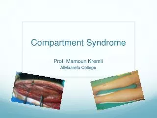
Compartment Syndrome A condition in which increased pressure within a limited space compromises the circulation and function of the tissues within that space.
Compartment SyndromeDefinition • Elevated tissue pressure within a closed fascial space • Reduces tissue perfusion - ischemia • Results in cell death - necrosis • True Orthopaedic Emergency
History • Volkmann 1881 • Richard von Volkmann published an article in which he attempted to describe the condition of irreversible contractures of the flexor muscles of the hand to ischemic processes occurring in the forearm • Application of restrictive dressing to an injured limb
History • Hildebrand 1906 • First used the term Volkmann ischemic contracture to describe the final result of any untreated compartment syndrome, and was the first to suggest that elevated tissue pressure may be related to ischemic contracture.
History • Thomas 1909 • Reviewed the 112 published cases of Volkmann ischemic contracture and found fractures to be the predominant cause. Also, noted that tight bandages, an arterial embolus, or arterial insufficiency could also lead to the problem
History • Murphy 1914 • First to suggest that fasciotomy might prevent the contracture. Also, suggested that tissue pressure and fasciotomy were related to the development of contracture
History • Ellis 1958 • Reported a 2% incidence of compartment syndrome with tibia fractures, and increased attention was paid to contractures involving the lower extremities
History • Seddon, Kelly, and Whitesides 1967 • Demonstrated the existence of 4 compartments in the leg and to the need to decompress more than just the anterior compartment. Since then, compartment syndrome has been shown to affect many areas of the body, including the hand, foot, thigh, and buttocks
Compartment SyndromeEtiology Compartment Size • tight dressing;Bandage/Cast • localised external pressure;lying on limb • Closure of fascial defects Compartment Content • Bleeding; Fx, vas inj, bleeding disorders • Capillary Permeability; • Ischemia / Trauma / Burns / Exercise / Snake Bite / Drug Injection / IVF
Fractures-closed and open Blunt trauma Temp vascular occlusion Cast/dressing Closure of fascial defects Burns/electrical Exertional states GSW IV/A-lines Hemophiliac/coag Intraosseous IV(infant) Snake bite Arterial injury Compartment SyndromeEtiology
Fracture • The most common cause • incidence of accompanying compartment syndrome of 9.1% • The incidence is directly proportional to the degree of injury to soft tissue and bone • occurred most often in association with a comminuted, grade-III open injury to a pedestrian Blick et al JBJS 1986
Blunt Trauma • 2nd most common cause • About 23% of CS • 25% due to direct blow McQueen et al; JBJS Br 2000
Incidence • McQueen et al; JBJS Br 2000 • 164 pts with CS, 149 male, 15 female • Most pts were usually under 35 • 69% with associated fx, about half were tibial shaft • 23% soft tissue injury without fx • Ranges of 2-12% have been published
Incidence McQueen et al; JBJS Br 2000
Patient positioning Meyer, Mubarak JBJS 2002
Patient Positioning • Leaving the calf free when the leg is placed in the hemilithotomy position instead of using a standard well-leg holder • Increases the difference between the diastolic blood pressure and the intramuscular pressure • May decrease the risk of compartment syndrome Meyer, Mubarak JBJS 2002
Compartment SyndromePathophysiology • Normal tissue pressure • 0-4 mm Hg • 8-10 with exertion • Absolute pressure theory • 30 mm Hg - Mubarak • 45 mm Hg - Matsen • Pressure gradient theory • < 20 mm Hg of diastolic pressure – Whitesides • McQueen, et al
Compartment SyndromeTissue Survival • Muscle • 3-4 hours - reversible changes • 6 hours - variable damage • 8 hours - irreversible changes • Nerve • 2 hours - looses nerve conduction • 4 hours - neuropraxia • 8 hours - irreversible changes
Compartment SyndromeDiagnosis • Pain out of proportion • Palpably tense compartment • Pain with passive stretch • Paresthesia/hypoesthesia • Paralysis • Pulselessness/pallor
Clinical Evaluation “Pain and the aggravation of pain by passive stretching of the muscles in the compartment in question are the most sensitive (and generally the only) clinical finding before the onset of ischemic dysfunction in the nerves and muscles.” Whitesides AAOS 1996
Clinical Evaluation • Pain – most important. Especially pain out of proportion to the injury (child becoming more and more restless /needing more analgesia) • Most reliable signs are pain on passive stretching and pain on palpation of the involved compartment • Other features like pallor, pulselessness, paralysis, paraesthesia etc. appear very late and we should not wait for these things. Willis &Rorabeck OCNA 1990
Clinical Evaluation • Beware of epidural analgesia • Strecker JBJS 1986 • Morrow J. Trauma 1994 • Beware long acting nerve blocks • Hyder JBJS Br 1995 • Beware controlled intravenous opiate analgesia
Compartment SyndromeDifferential Diagnosis • Arterial occlusion • Peripheral nerve injury • Muscle rupture
Compartment SyndromePressure Measurements • Suspected compartment syndrome • Equivocal or unreliable exam • Clinical adjunct • Contraindication • Clinically evident compartment syndrome
Infusion manometer saline 3-way stopcock (Whitesides, CORR 1975) Catheter wick slit wick Arterial line 16 - 18 ga. Needle (5-19 mm Hg higher) transducer monitor Stryker device Side port needle Compartment SyndromePressure Measurements
Compartment SyndromePressure Measurements • Arterial line • Zero at the level of the affected limb
Compartment SyndromePressure Measurements • Simple Needle • 18 gauge • Least accurate • Usually gives falsely higher reading • Slit Catheter and Side ported needle • No significant difference • More accurate Side port Moed et al JBJS 1993
Compartment SyndromePressure Measurements • Measurements must be made in all compartments • Anterior and deep posterior are usually highest • Measurement made within 5 cm of fx • Marginal readings must be followed with repeat physical exam and repeat compartment pressure measurement Heckman, WhitesidesJBJS 1994
Unequivocal + Findings FASCIOTOMY Pt. not alert/polytrauma/inconc. Comp. pressure measurement w/i 30 mm Hg >30 mm Hg of DBP Serial exams FASCIOTOMY SUSPECTED COMPARTMENT SYNDROME McQueen JBJSB 1996
Threshold for fasciotomy • McQueen, Court-Brown JBJS Br 1996 • 116 pts with tibial diaphyseal fx had continuous monitoring of anterior compartment pressure for 24 hours • 53 pts had ICP over 30 mmHg • 30 pts had ICP over 40 mmHg • 4 pts had ICP over 50 mmHg • Only 3 had delta pr(DBP-ICP) of < 30, they had fasciotomy • None of the patients had any sequelae of the compartment syndrome • Decompression should be performed if the differential pressure level drops to under 30 mmHg
Medical Management • Ensure patient is normotensive ,as hypotension reduces prefusion pressure and facilitates further tissue injury. • Remove cicumferential bandages and cast • Maintain the limb at level of the heart as elevation reduces the arterial inflow and the arterio-venous pressure gradient on which perfusion depends. • Perfusion pressure = A pr(30-35mmHg) – V pr(10-15mmHg) • Supplemental oxygen administration.
Medical Management • Compartmental pressure falls by 30% when cast is split on one side • Falls by 65% when the cast is spread after splitting. • Splitting the padding reduces it by a further 10% and complete removal of cast by another 15% • Total of 85-90% reduction by just taking off the plaster! Garfin, Mubarak JBJS 1981
Compartment SyndromeEmergent Treatment • Remove cast or dressing • Place at level of heart (DO NOT ELEVATE to optimize perfusion) • Alert OR and Anesthesia • Bedside procedure • Medical treatment
Surgical Treatment • Fasciotomy, Fasciotomy, Fasciotomy, • All compartments !!!
Compartment SyndromeSurgical Treatment • Fasciotomy - prophylactic release of pressure before permanent damage occurs. Will not reverse injury from trauma. • Fracture care – stabilization • Ex-fix • IM Nail
Compartment SyndromeIndications for Fasciotomy • Unequivocal clinical findings • Pressure within 15-20 mm hg of DBP • Rising tissue pressure • Significant tissue injury or high risk pt • > 6 hours of total limb ischemia • Injury at high risk of compartment syndrome • CONTRAINDICATION - Missed compartment syndrome (>24-48 hrs)
Fasciotomy Principles • Make early diagnosis • Long extensile incisions • Release all fascial compartments • Preserve neurovascular structures • Debride necrotic tissues • Coverage within 7-10 days
Compartment SyndromeLower Leg • 4 compartments • Lateral: Peroneus longus and brevis • Anterior: EHL, EDC, Tibialis anterior, Peroneus tertius • Supeficial posterior-Gastrocnemius, Soleus • Deep posterior-Tibialis posterior, FHL, FDL • .
Single Incision • Perifibular Fasciotomy • Matsen et al (1980) • Single incision just posterior to fibula • Common peroneal nerve
Double Incision • In most instances it affords better exposure of the four compartments • 2 vertical incisions separated by minimum 8 cm • One incision over anterior and lateral compartments • Superficial peroneal nerve • One incision located 1-2 cm behind postero-medial aspect of tibia • Saphenous nerve and vein Mubarak et al JBJS 1977
Fasciotomy: Medial Leg Gastroc-soleus Flexor digitorum longus
Fasciotomy: Lateral Leg Intermuscular septum Superficial peroneal nerve
Look for Superficial Peroneal Nerve • superficial peroneal nerve exits from lateral compartment about 10 cm above lateral malleolus and courses into the anterior compartment • Risk of injury
Use a Generous Incision • Lengthening the skin incisions to an average of 16 cm decreases intracompartmental pressures significantly. • The skin envelope is a contributing factor in acute compartment syndromes of the leg and The use of generous skin incisions is supported Cohen, Mubarak JBJS Br 1991
Compartment SyndromeForearm • Anatomy-3 compartments • Mobile wad-BR,ECRL,ECRB • Volar-Superficial and deep flexors • Dorsal-Extensors • Pronator quadratus described as a separate compartment
Forearm Fasciotomy • Volar-Henry approach • Include a carpal tunnel release • Release lacertus fibrosus and fascia • Protect median nerve, brachial artery and tendons after release
Forearm Fasciotomy • Protect median nerve, brachial artery and tendons after release • Consider dorsal release
Compartment SyndromeFoot • 9 compartments • Medial, Superficial, Lateral, Calcaneal • Interossei(4), Adductor • Careful exam with any swelling • Clinical suspicion with certain mechanisms of injury • Lisfranc fracture dislocation • Calcaneus fracture