Download
1 / 65
650 likes | 652 Vues
Lymphoid System I & II. Dr. Jack L. Haar Department of Anatomy and Neurobiology. Overview of Immune System. Functions Provides immune surveillance and defense Provides immune tolerance (distinguishes self from non-self) Absorbs lipids

E N D
Lymphoid System I & II Dr. Jack L. Haar Department of Anatomy and Neurobiology
Overview of Immune System • Functions • Provides immune surveillance and defense • Provides immune tolerance (distinguishes self from non-self) • Absorbs lipids • Maintains fluid balance by returning tissue fluid and lymphocytes to the blood
Overview continued • Concept of immunity • Immune system provides a way to recognize “self” from “non-self” • An individual mounts an immune response to foreign material • If person survives an attack by foreign material (infection), immunity may result, meaning the foreign material is “remembered” • Thus immunity has specificity and memory
Overview continued • Terminology • Antigen: a foreign (non-self) substance. Could be bacterial, tumor cells, transplanted cells or virus-infected cells • Antibody: circulating protein in blood plasma (immunoglobulin) that interacts with a specific antigen • Humoral immunity: antibodies against antigen circulating in blood stream; produced by plasma cells derived from B lymphocytes • Cellular (cell-mediated) immunity: Immunocompetent cells contact, react against, and destroy antigen; mediated by T lymphocytes
Cells of the Immune response20 – 50% WBC’s are lymphocytes
A. 35% of circulating lymphocytes are B lymphocytes or B cells Become Plasma cells or B memory cellsB. 65% of circulating lymphocytes are T lymphocytes or T cells Become Helper, Killer, or T memory cells
ImmunoblastsForm from T or B lymphocytes when stimulated by Antigen • Antigen -presenting cellsMonocyte-macrophage derived. Process antigen to present to lymphocytes • Includes macrophages, epidermal Langerhans cells, dendritic cells of lymphoid organs, and epithelial cells of thymus
Thymus undergoes involution with age • Is influenced by adrenal cortical steroids and radiation • Thymus is a bilobed organ
Stroma formed from thymic epithelial cells with bundles of tonofibrils and linked by desmosomes • Thymocytes develop from HSC from yolk sac (mesoblastic), fetal liver (hepatic), and bone marrow (myeloid) phases of development • Thymus is not exposed to external environment
Histology • Dense irregular CT capsule • Septa form lobules • Cortex and medulla • Stroma formed from thymic epithelial cells
Cortex • Thymic epithelial cells • Large numbers of thymocytes (lymphocytes) • Macrophages
Medulla • Thymocytes are larger and fewer in number than in cortex Only 5% of thymocytes are in the medulla • Hassall’s corpuscles form from the stromal cells Keratohyaline granules
Vessels enter and leave through the capsule and follow the septa • Blood thymus barrier
Function of the thymus • Thymus is seeded with HSC’s • Thymocytes proliferate in cortex • Self-reactive thymocytes are eliminated • Non-self reactive cells migrate to medulla • Cells enter blood stream and migrate to secondary lymphoid organs • They occupy T-dependant areas of lymphoid organs where they nest, divide, and mature • The thymus is an endocrine organ and responds to many hormones, viz. ACTH, GH, and sex hormones • Thymectomy at birth impairs immune function
Bursa of Fabricius • Functions in birds as primary lymphoid organ giving rise to B-lymphocytes
Bone marrow • Functions in mammals as primary lymphoid organ giving rise to B-lymphocytes
MALT • GI tract • Respiratory tract • Genito-urinary tract • Diffuse lymphatic tissue • Lamina propria • Reticular fibers • Lymphocytes • Primary lymphatic nodule
Secondary Lymphatic nodule • Germinal center • B-dependant spherical area • Cap of tightly packed small lymphocytes • Not encapsulated by C.T.
Germinal center • Oval pale-staining area • Contains dividing cells and macrophages
Solitary nodules • Not encapsulated by C.T. • May be primary or secondary
High endothelial venules • Associated with diffuse lymphatic tissue • Allow lymphocytes to escape from vessel
Locations of aggregates of nodules • Peyer’s patches • Typhoid nodules • Appendix
Typhoid Fever - a bacterial illness caused by Salmonella typhi
Lymphoid System II Dr. Jack L. Haar Department of Anatomy and Neurobiology
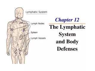
Lymphatic Vessels • Lymphatic capillaries • Lacteals for lipid • Valves direct flow • Asymmetrical system • Edema
Lymphatic filariasis, also known as elephantiasis, is best known from dramatic photos of people with grossly enlarged or swollen arms and legs. The disease is caused by parasitic worms, including Wuchereria bancrofti, Brugia malayi, and B. timori, all transmitted by mosquitoes. Lymphatic filariasis currently affects 120 million people worldwide, and 40 million of these people have serious disease.
Lymph node Histology • Distributed along vessels • Filter lymph • Produce lymphocytes • Dense CT capsule • Afferent lymphatics on convex surface • Reticular fiber framework Subcapsular sinus Lymphoid nodule in cortex
Hilus • Blood vessels enter and leave • Efferent lymphatics leave node
Cortex of lymph node • Lymphoid nodules and diffuse lymphatic tissue • Lymphatic sinuses • Subcapsular • Cortical
Medulla of lymph node • Medullary cords • Medullary sinuses
Blood vessels follow trabeculae into nodeHEV with cuboidal endothelium Lymphocytes pass from blood into lymph node