Download
1 / 15
150 likes | 327 Vues
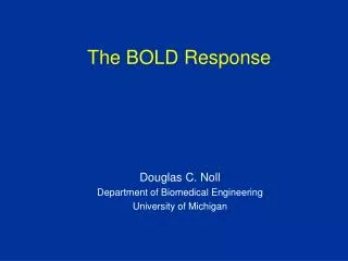
Motor Inhibition and the BOLD response in the primary motor cortex. Archana Purushotham, James Ashe & Seong-Gi Kim Center for Magnetic Resonance Research, Departments of Biomedical Engineering and Neuroscience, University of Minnesota. Introduction.

E N D
Motor Inhibition and the BOLD response in the primary motor cortex Archana Purushotham, James Ashe & Seong-Gi Kim Center for Magnetic Resonance Research, Departments of Biomedical Engineering and Neuroscience, University of Minnesota
Introduction Understanding the neural correlates of BOLD activity: • What do ‘hot spots’ on fMRI maps mean? • Does the BOLD signal result from excitation alone, or from excitation as well as inhibition?
Hot spots on fMRI maps currently interpreted as functionally excited areas, with neural spiking output to other areas – typically assumed to be mutually connected to form a functional neural circuit. • Neural inhibition thought not to evoke a BOLD response(Waldvogel et al., 2000)
Energy consumption modulates CBF and the BOLD signal (Lauritzen, 2001; Arthurs and Boniface, 2002; Logothetis et al., 2001; Raichle, 2001); both synaptic excitation and inhibition consume energy • Glucose consumption (Ackermann et al., 1984; Nudo and Masterton, 1986) and CBF (Mathiesen et al., 1998) increase during inhibition • BOLD signal correlates most closely with local field potentials – however, study primarily of excitatory activity (Logothetis et al., 2001 ) • => Neural inhibition would also be expected to cause a BOLD response from a metabolic point of view
Neural inhibition and the No-go response in the primary motor cortex • TMS studies of Go/No-go tasks (Waldvogel et al., 2000; Sohn et al., 2002): Inhibition of primary motor cortex during the No-go condition • No-go related local field potential changes in the primary motor cortex (direct recording - Sasaki and Gemba, 1986)
Given that the primary motor cortex is functionally inhibited, with no output to the pyramidal tract during No-go trials (cancellation of an intended movement), is there a corresponding BOLD response?
Paradigm A delayed, cued Go/No-go joystick task: GO delay (0-7000 ms) movement control 200 ms 1600 ms NO-GO control 7300 ms • Variable, pseudo-randomized delay periods : 0, 2, 4 and 7 sec • No-go trials constitute 20% of all trials • Trial epochs : 30 or 35 seconds long; 30-40 trials
Data Acquisition fMRI • Single-shot 64 x 64 EPI images using a 4 Tesla MRI system • 5 axial sections 5 mm thick, including the primary (M1), supplementary (SMA) and pre-SMA • TR = 1 s for 9 subjects; 0.5 s for 5 subjects • Structural images 128 x 128 T1-weighted (FLASH) • 14 normal adult, right-handed human subjects Electromyography • Surface EMG of flexors and extensors of forearm, while imaging in 3 subjects, only pre-fMRI training for 1.
Data Analysis • Temporal smoothing, discarded errors • fMRI epochs grouped by delay period and averaged to get mean timecourse for each delay condition; linearly detrended • M1, SMA and pre-SMA demarcated manually • Active voxels chosen based on direct movement time-course using cross-correlation (alpha = 0.01) • Epoch time-courses for each delay condition created by averaging over activated voxels in each area • Data from subjects with poor signal-to-noise ratio (1) and absence of No-go activity in pre-SMA (2) excluded
* Results Mean reaction time Reaction time (ms) Delay (s)
go no-go Pooled (8 subjects) data Pre-SMA cM1 Pre-SMA cM1 BOLD signal change (%) Time (s)
go no-go Individual data (3/8) Pre-SMA cM1 BOLD signal change (%) Time (s)
In spite of the known functional inhibition of the primary motor cortex during the No-go condition, there is a significant increase in the BOLD signal.
Conclusions • A BOLD response can occur in a functionally inhibited cortical region. • Hot spots on fMRI brain maps do not automatically imply an increase in spike output from that area of the brain, nor that these spots form a functionally connected neuronal circuit.
The BOLD response appears to correlate better with LFP (summated synaptic activity), rather than action potentials, even in a situation where inhibitory neural interactions are prominent. • This supports the prediction based on energetics, that neural inhibition also causes a BOLD response, similar to neural excitation.