Download
1 / 87
940 likes | 1.01k Vues
Neuraxial Blokade. Dr: Ahmed Thallaj Associate Professor, college of medicine, KSU Head of regional Anesthesia division. Objectives. Relevant anatomy and surface landmark for Neuraxial block. Differences between spinal and epidural. Equipment and local anesthetics.

E N D
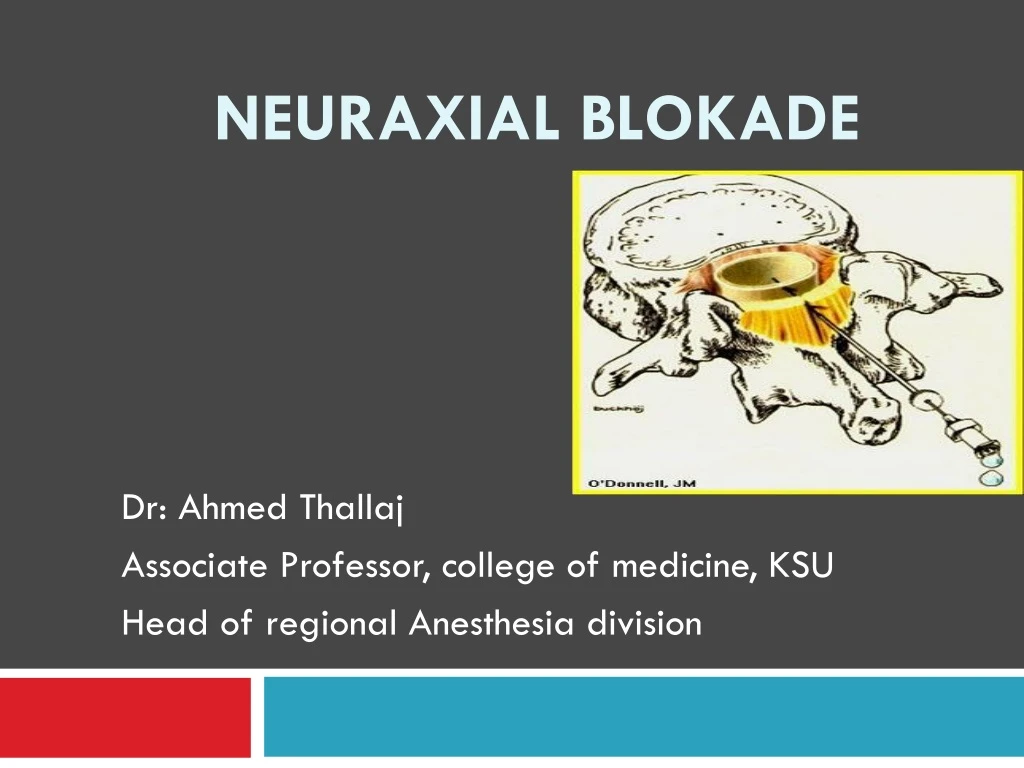
NeuraxialBlokade Dr: Ahmed Thallaj Associate Professor, college of medicine, KSU Head of regional Anesthesia division
Objectives • Relevant anatomy and surface landmark for Neuraxial block. • Differences between spinal and epidural. • Equipment and local anesthetics. • Indication and contraindication. • Side effects, complications and treatment. • LAST
Knowledge of anatomy for neuraxial blockade is essential! • 7 cervical vertebrae • 12 thoracic vertebrae • 5 lumbar vertebrae • Sacrum • Coccyx
Cervical Vertebrae Thoracic Vertebrae Lumbar Vertebrae
Individual Vertebral Anatomy • Each vertebra consists of a pedicle, transverse process, superior and inferior articular processes, and a spinous process. • Each vertebra is connected to the next by intervertebral disks. • There are 2 superior and inferior articular processes (synovial joints) on each vertebra that allows for articulation. • Pedicles contain a notch superiorly and inferiorly to allow the spinal nerve root to exit the vertebral column.
Vertebral Anatomy- Top View Spinous Process Transverse Process Lamina Spinal Canal Vertebral Body
Intervertebral Disc Intervertebral Foramina Spinal Nerve Root
Thoracic Vertebrae Angle of transverse process will affect how the needle is orientated for epidural anesthesia or analgesia. With flexion the spinous process in the lumbar region is almost horizontal. In the thoracic region the spinous process is angled in a slight caudad angle. Lumbar Vertebrae
L 2 L 5 Interlaminar spaces are larger in the lower lumbar region. If an anesthesia provider finds it challenging at one level it is important to remember that moving down one space may provide a larger space.
Ligaments that support the vertebral column Ventral side: Anterior and posterior longitudinal ligaments Dorsal side: Important since these are the structures your needle will pass through!
Ligaments are identified by tactile sensation (feel) Dorsal ligaments transversed during neuraxial blockade. With experience the anesthesia provider will be able to identify anatomical structures by “feel”.
Termination of Spinal Cord In adults usually ends at L1. Infants L3 There are anatomical variations. For most adults it is generally safe to place a spinal needle below L2.
Locating prominent cervical and thoracic vertebrae • C2 is the first palpable vertebrae • C7 is the most prominent cervical vertebrae • With the patients arms at the side the tip of the scapula generally corresponds with T7
Spinous Processes • Generally are palpable to help identify the midline • If unable to palpate the spinous process one can look at the upper crease of the buttocks and line up the midline as long as there is no scoliosis or other deformities of the spine
What is Tuffier’s Line? • A line drawn between the highest points of both iliac crests will yield either the body of L4 or the L4-L5 interspace.
Anatomical Considerations of the Spinal Cord and Neuraxial Blockade.
The Subarachnoid Space is a continuous space that contains • CSF • Spinal cord & nerves
CSF • Clear fluid that fills the subarachnoid space • Total volume in adults is ~100-150 ml (2 ml/kg) • Volume found in the subarachnoid space is ~35-45 ml • Continually produced at a rate of 450 ml per 24 hour period replacing itself 3-4 times
CSF • Reabsorbed into the blood stream by arachnoidvilli. • Specific gravity is between 1.003-1.007 (this will play a crucial role in the baracity of local anesthetic that one chooses) • CSF plays a role the patient to patient variability in relation to block height and sensory/motor regression (80% of the patient to patient variability) • Body wt is the only measurement that coincides with CSF volume (this becomes important in the obese and pregnant).
Membranes that surround the spinal cord • Pia mater- highly vascular, covers the spinal cord and brain, attaches to the periosteum of the coccyx ( Filumterminalis) • Arachnoid mater- non vascular and attached to the dura mater. Principal barrier to the migration of medications in and out of the CSF. • Dura mater (“tough mother”)- extension of the cranial dura mater, extends from the foramen magnum to S2.
Filum Terminale • An extension of the pia mater that attaches to the periosteum of the coccyx.
Epidural Space Anatomy • Extends from the formen magnum to the sacral hiatus
Epidural Space Anatomy • The epidural space surrounds the dura mater anteriorly, laterally, and most importantly to us posteriorly.
The Bounds of the Epidural Space are as follows: • Anterior- posterior longitudinal ligament • Lateral- pedicles and intervertebral ligaments • Posterior- ligamentum flavum
Ligamentum Flavum • Posterior to the epidural space • Extends from the foramen magnum to the sacral hiatus • Distance from skin to ligament varies from 3-8 cm in the lumbar area. It is 4 cm in 50% of the patients and 4-6 cm in 80% of the patients. • Thickness of the ligamentumflavum also varies. In the thoracic area it can range from 3-5 mm and in the lumbar it can range from 5-6 mm
Contents of the Epidural Space • Fat • Areolar tissue • Lymphatics • Blood vessels including the Batson venous plexus
Spinal anesthesia : Injection of small amounts ( 2-3 ml) of local anaesthetics into the CSF at the level below ( L2 ) ,where the spinal cord ends, anesthesia of the lower body part below the umbilicus is achieved. Indication Operations below the umbilicus: hernia repairs, gynaecological, urological operation, orthopedics, Any operation on the perineum or genitalia. Definition
Spinal Anesthesia • Contraindications • Absolute: • Refusal • Infection • Coagulopathy & anticoagulated patient • Severe hypovolemia • Increased intracranial pressure • Severe aortic or mitral stenosis • Relative: • Use your best judgment
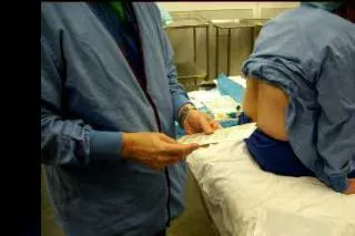
Spinal Technique • Midline Approach • Skin • Subcutaneous tissue • Supraspinous ligament • Interspinous ligament • Ligamentumflavum • Epidural space • Dura mater • Arachnoid mater • Paramedian or Lateral Approach • Same as midline excluding supraspinous & interspinous ligaments
PDPH • Develop 12-48 hours after spinal anesthesia. • Headache improve when lying supine.