Download
1 / 45
470 likes | 791 Vues
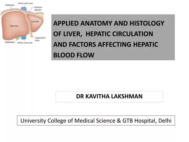
APPLIED ANATOMY AND HISTOLOGY OF LIVER , HEPATIC CIRCULATION AND FACTORS AFFECTING HEPATIC BLOOD FLOW. dR kavitha lakshman. University College of Medical Science & GTB Hospital, Delhi. ANATOMY OF THE LIVER. Largest gland, largest reticuloendothelial organ

E N D
APPLIED ANATOMY AND HISTOLOGY OF LIVER, HEPATIC CIRCULATION AND FACTORS AFFECTING HEPATIC BLOOD FLOW dRkavithalakshman University College of Medical Science & GTB Hospital, Delhi
ANATOMY OF THE LIVER • Largest gland, largest reticuloendothelial organ • Weighs around 1.2 to 1.6 Kg • Occupies the right upper quadrant of the abdomen • Invested in peritoneum except – gallbladder bed, IVC, bare area of liver • Ligaments- duplication of peritoneum over the liver
DIVISIONS OF THE LIVER • Falciform ligament-oversimplified,anatomically incorrect • Cantlie's line- a plane running from the gall-bladder to the left side of the IVC • Couinad’s segments- 8 functional segments, each supplied by a pedicle • Bismuth’s classification- 4 sectors separated by scissurae containing the three main Hepatic vein
NERVE SUPPLY AND LYMPHATIC • Sympathetic nerve fibers - T6–T11 Parasympathetic fibers - Rt&Ltvagus RtPhenic.N • Superficial lymphatics:Surface of organ and terminate in Caval ,Hepatic,Celiac lymph nodes • Deep lymphatics: IVC and Hepatic nodes
HEPATOCYTES Hepatocytes - polyhedral , with a central spherical nucleus. Single-cell-layer plates Lined on either sides by sinusoids. Every hepatocyte has contact with adjacent hepatocytes, the biliary space (bile canaliculus), and the sinusoidal space, allowing its broad range of functions
SINUSOIDS • Sinusoids are blood filled spaces • 10um in diameter • Hepatic and Portal blood mixes here • Endothelial cells -lack intercellular junctions -Absence of basement membrane -Contain multiple and large fenestrations. Maximal contact of hepatocyte membrane(space of Disse) and blood in the sinusoidal space. Permits free bidirectional movement of solutes
KUFFER CELLS • Resident macrophages line sinusoids • Partly derived from bone marrow monocytes • Clearance of gut derived toxins • Engulf debris and dead rbcs • Secrete cytokines IL-1,IL-6,TNF alfa • Express class II MHC- antigen presentation
ITO CELLS • Found in the space of Disse. • They have dendritic processes which are in contact with hepatocytemicrovilli and endothelial cells. • Vitamin A storage • Synthesis of extracellular collagen. • Play a central role in the development and progression of hepatic fibrosis
HEPATIC LOBULE 50,000-100000 lobules Functional unit of the liver
FUNCTIONS OF HEPATOCYTES Metabolism of proteins, carbohydrates, lipids Metabolism of heme, bile Synthesis of coagulants-2,7,9,10,protein S,C Drug metabolism Immune defense
DRUG METABOLISM Phase 1 Metabolism e.g. oxidations, reductions, hydrolysis convert drugs to more polar compounds The products of phase 1 metabolism are -Readily excreted in bile or urine than their precursors are. -These products may also be substrates for phase 2 conjugations.
Phase 2 metabolism Conjugate xenobiotics or their metabolites with endogenous hydrophilic molecules such as glucuronic acid, acetate, sulfates, amino acids, and glutathione, glucuronic acid etc When compared with their precursors, conjugated xenobiotics are usually less efficacious, less toxic, more hydrophilic, and more readily excreted in bile or urine.
Phase 3 Elimination • Involve specific molecular transporters—known as ATP-binding cassette (ABC) transport proteins—that facilitate the excretion of xenobiotics and endogenous compounds. • These proteins use ATP hydrolysis to drive molecular transport. • Major ABC transport proteins include cystic fibrosis transmembrane conductance regulator (CFTR), canalicular copper transporters, and multidrug resistance protein (MDR).
dysfunction of ABC transport proteins can disrupt the flow of bile, impair excretion of xenobiotics and endogenous compounds, and induce cholestatic liver injury.
Blood supply of liver The liver is at the hub of the splanchnic circulation. Receives 25% of the total cardiac output via a dual vascular supply. Arterial blood is supplied via the hepatic artery—a branch of the common hepatic artery that has its origin at the celiac trunk of the abdominal aorta On the other hand, the portal vein has as its tributaries the superior mesenteric and splenic veins, which carry the entire venous drainage of the preportalsplanchnic beds.
HA+PV HEPATIC ARTERIOLE+PORTAL VENULE SINUSOIDS(FENESTERATED ENOTHELIUM) HEPATOCYTES SINUSOIDS SPACE OF DISSE CENTRAL VEIN(TERMINALHEPATIC VENULE) LOBULE MAIN HEPATIC VEIN IVC
PORTAL VEIN • Begins and ends in a network of capillaries • lack of valves - accommodate high flow at low pressure because of the low resistance • 75% of hepatic blood flow • Postcapillary and largely deoxygenated • large-volume flow rate provides 50% to 70% of the liver's oxygenation.
Numerous connections exists between the portal venous system and the systemic venous system. • Provide collateral supply under conditions of high portal venous pressure 1. Submucosal veins of the proximal stomach and distal esophagus, which receive portal flow from the short gastric veins and the left gastric vein 2.Umbilical and abdominal wall veins, which recanalize from flow through the umbilical vein in the ligamentumteres 3.Superior hemorrhoidal plexus, which receives portal flow from inferior mesenteric vein tributaries
FACTORS AFFECTING THE HEPATIC BLOOD FLOW INTRINSIC REGULATION • HEPATIC ARTERIAL BUFFER RESPONSE • METABOLIC CONTROL • PRESSURE FLOW AUTO REGULATION EXTRINSIC REGULATION • NEURAL CONTROL • HUMORAL CONTROL
HEPATIC ARTERIAL BUFFER RESPONSE • Change in portal venous flow-reciprocal change in hepatic arterial flow • Adenosine in periportal region • Can double hepatic arterial flow • Disrupted in endotoxemia , splancnichypoperfusion, portal inflow falls by >50%
PRESSURE FLOW AUTOREGULATION • Myogenic response –vascular smooth muscle • Transmural pressure-myogenic tone-vessel caliber-dec blood flow • Hepatic artery-present • Fed state- active • Is of minimal significance intraoperatively
METABOLIC CONTROL • Low pH and oxygen tension portal blood increases hepatic blood flow • Postprandial hyperosmolarity increases both hepatic and portal blood flow
NEURAL CONTROL • Post ganglionic sym. T6-T11 Vagus ,Phrenic nerve • Sympathoadrenal stimulation (hypercarbia,hypoxia,pain) vasoconstriction—decrease in hbf • With in sec, splanchnic stimulation autotransfuse 400-500 ml (80% total hepatic blood vol)into central circulation in healthy euvolemic individuals
HUMORAL CONTROL • EPINEPHRINE Hepatic a.— vasoconstriction followed by vasodilation Portal v--- vasoconstriction • DOPAMINE -vasodilation • Glucagon -vasodilation • Angitention II -vasoconstriction • Vasopressin - constricts splanchic arterial bed decreases portal venous resistance
FACTORS INCREASING HEPATIC BLOOD FLOW FACTORS DECREASING HEPATIC BLOOD FLOW IPPV+PEEP SURGERY HYPOCAPNIA;HYPOXIA UPRIGHT POSTURE CIRRHOSIS DRUGS A-AGONIST B-BLOCKERS H2 BLOCKERS CIMETIDINE,RANITIDINE,VASOPRESSIN ANESTHETICS-VOLATILE AND INTRAVENOUS • HYPERCAPNIA • AC.HEPATITIS -VIRAL,ALCOHOLIC • SUPINE POSTION • FOOD • DRUGS B-AGONIST PHENOBARBITONE
EFFECT OF ANAESTHESIA ON HEPATIC BLOOD FLOW • Hepatic blood flow usually decreasesduring regional and general anesthesia • All volatile anesthetic agents reduce portal hepatic blood flow- halothane • Indution agents- ketamine increases flow • GA-decrease in map, co, reflex sympathetic stimulation • Spinal and epidural anesthesia decrease hepatic blood flow primarily by lowering arterial blood pressure.
Ventilation- IPPV, PEEP Surgery – direct vascular compression, local reflexes, reflex sympathetic stimulation
REFERENCES • PHILIP SM, SIMON G, HEPATIC PHYSIOLOGY AND PATHOLOGY,MILLERS 7TH EDITION,411-435 • BRAIN S, DAVID ROCCAFORTE. HEPATIC ANATOMY, FUNCTION AND PHYSIOLOGY. CLINICAL ANAESTHESIA,6TH EDITION,1247-78 • EDWARD MORGAN . HEPATIC PHYSIOLOGY AND ANAESTHESIA.CLINICAL ANAESTHESIOLOGY, 4TH EDITION • INTERNATIONAL PRACTICE OF ANAESTHESIA, PRYS ROBERTS • MICHAEL A, YUMNG F, ANATOMY AND PHYSIOLOGY OF THE LIVER,SABISTON TEXT BOOK OF SURGERY, 18TH EDITION • NEIL R B,ANATOMY OF THE LIVER .GRAY’S ANATOMY 39TH EDITION • HEPATIC PHYSIOLOGY,GANONG 22ND EDITION • STOELTING R, HILLIER S, LIVER AND GASTROINTESTINAL TRACT PHARMACOLOGY AND PHYSIOLOGY IN ANAESTHETIC PRACTICE, 4TH EDITION