Download
1 / 36
360 likes | 365 Vues
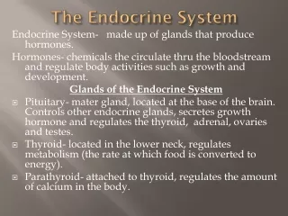
PART 2. The Endocrine System. The Parathyroid Gland. Lie on the posterior surface of the thyroid gland Contain two types of endocrine cells Chief cells Produce parathyroid hormone (PTH) Increases blood concentration of Ca 2+ Oxyphil cells Function unknown. The Parathyroid Gland.

E N D
PART 2 The Endocrine System
The Parathyroid Gland • Lie on the posterior surface of the thyroid gland • Contain two types of endocrine cells • Chief cells • Produce parathyroid hormone (PTH) • Increases blood concentration of Ca2+ • Oxyphil cells • Function unknown
The Parathyroid Gland Figure 25.7a
The Parathyroid Gland Figure 25.7b, c
The Adrenal Glands • Located on the superior surface of the kidneys • Nerve supply is almost exclusively sympathetic fibers • Two endocrine glands in one • Adrenal medulla – a knot of nervous tissue • Adrenal cortex – bulk of the adrenal gland
The Adrenal Medulla • Discussed in Chapter 15 – the ANS • Chromaffin cells • Modified postganglionic sympathetic neurons • Secrete catecholamines • Active in “fight, flight, and fright” response
The Adrenal Cortex • Secretes a variety of hormones • All are steroids • Cortex is composed of three layers • Zona glomerulosa • Zona fasciculata • Zona reticularis
The Adrenal Gland Figure 25.8a, b
Adrenal Corticosteroids • Two main classes • Mineralocorticoids • Aldosterone is secreted by the zona glomerulosa • Glucocorticoids • Cortisol • Secreted by zona fasciculata and zona reticularis • Helps the body deal with stressful situations • Dehydroepiandrosterone (DHEA)
Structure of Steroid-Secreting Cells • Includes cells of • Adrenal cortex • Testicular and ovarian cells
Structure of Steroid-Secreting Cells • Steroid-secreting cells have distinctive features • Abundant smooth ER and no secretory granules • Mitochondria have unusual cristae • Shaped like tubes • Lipid droplets are abundant in cytoplasm • Lipids are the raw material of steroids
Steroid-Secreting Hormones of Male Figure 25.9
The Pineal Gland • Located on the roof of the diencephalon • Shaped like a pinecone • “Pineal sand” is radiopaque • Used as a landmark to identify other brain structures in X-Rays • Pinealocytes secrete melatonin • A hormone that regulates circadian rhythms
The Pancreas • Located in the posterior abdominal wall • Contains endocrine and exocrine cells • Exocrine cells • Acinar cells – secrete digestive enzymes • Endocrine cells • Pancreatic islets – islets of Langerhans • About one million islets – scattered throughout the pancreas
The Pancreas • Main endocrine cell types • Alpha cells ( cells) – secrete glucagon • Signals liver to release glucose from glycogen • Raises blood sugar • Beta cells ( cells) – secrete insulin • Signals most body cells to take up glucose from the blood • Promotes storage of glucose as glycogen in liver • Lowers blood sugar
The Thymus • Located in the lower neck and anterior thorax • Important immune organ • Site at which T-lymphocytes arise from precursor cells
The Gonads • Main sources of sex hormones • Testes and ovaries • Male • Interstitial cells secrete androgens • Primarily testosterone • Promotes the formation of sperm • Maintains secondary sex characteristics PLAY Male Hormones
The Gonads • Female • Ovaries • Androgens secreted by the theca folliculi • Converted to estrogen by follicular granulosa cells • Estrogen • Maintains secondary sex characteristics • Progesterone • Prepares the uterus for pregnancy
Other Endocrine Structures • Endocrine cells occur within • The heart • Atria contain atrial natriuretic peptide (ANP) • The GI tract • Enteroendocrine cells • The placenta • Sustains the fetus and secretes several steroid protein hormones
Other Endocrine Structures • The kidneys • Cells of the juxtaglomerular apparatus (JGA) secrete renin • Endothelial cells and interstitial connective tissue – secrete erythropoietin • The skin • Modified cholesterol molecules convert to a precursor of vitamin D
Pituitary Disorders • Gigantism • Hypersecretion of GH in children • Pituitary dwarfism • Hyposecretion of GH • Diabetes insipidus • Pars nervosa does not make enough ADH
Disorders of the Pancreas: Diabetes Mellitus • Caused by • Insufficient secretion of insulin • Resistance of body cells to the effects of insulin • Type 1 diabetes • Develops suddenly, usually before age 15 • T cell-mediated autoimmune response destroys beta cells
Diabetes Mellitus • Type 2 diabetes • Adult onset • Usually occurs after age 40 • Cells have lowered sensitivity to insulin • Controlled by dietary changes and regular exercise
Disorders of the Thyroid Gland • Grave’s disease • Most common type of hyperthyroidism • Immune system makes abnormal antibodies • Stimulates the oversecretion of TH by follicle cells • Leads to nervousness, weight loss, sweating, and rapid heart rate
Disorders of the Thyroid Gland • Myxedema • Adult hypothyroidism • Antibodies attack and destroy thyroid tissue • Low metabolic rate and weight gain are common symptoms
Disorders of the Thyroid Gland • Endemic goiter • Due to lack of iodine in the diet • Cretinism • Hypothyroidism in children • Short, disproportionate body, thick tongue and mental retardation
Thyroid Disorders Figure 25.11
Disorders of the Adrenal Cortex • Cushing’s syndrome • Caused by hypersecretion of glucocorticoid hormones – usually a pituitary tumor • Addison’s disease • Hyposecretory disorder of the adrenal cortex • Deficiencies of both mineralocorticoids and glucocorticoids
Thyroid Disorders Figure 25.12
Embryological Origin of Selected Endocrine Organs • Thyroid gland • Forms from a thickening of endoderm on the floor of the pharynx • Parathyroids and the thymus gland • From endoderm lining the pharyngeal pouches
Embryological Origin of Selected Endocrine Organs • Pineal gland • Originates from ependymal cells • Pituitary gland – dual origin • Adenohypophysis originates from the roof of the mouth • Neurohypophysis grows inferiorly from the floor of the brain
Embryological Origin of Selected Endocrine Organs • Adrenal gland – dual origin gland • Adrenal medulla – from neural crest cells of nearby sympathetic trunk ganglia • Adrenal cortex – from mesoderm lining the coelom
The Endocrine System Throughout Life • Endocrine organs operate effectively until old age • Adenohypophysis • Increase in connective tissue and lipofuscin • Decrease in vascularization and number of hormone-secreting cells • Adrenal cortex • Normal rates of glucocorticoid secretion continue • Adrenal medulla • No age-related changes in catecholamines
Embryonic Development of Some Major Endocrine Organs Figure 25.13a, b
Embryonic Development of Some Major Endocrine Organs Figure 25.13c, d
The Endocrine System Throughout Life • Thyroid hormones • Decrease slightly with age • Parathyroid glands • Little change with aging • GH, DHEA, and the sex hormones • Marked drops in secretion with age