Download

1 / 22
220 likes | 361 Vues
Identifying the Mechanical Properties of Biological Materials 6th November 2008 ARUP Campus, Solihull. CHANGES IN CHARACTERIZATION OF THE HUMAN SKIN DUE TO THE PROCESS OF SUCCESSIVE SKIN EXPANSION: an in vivo characterization using Delfino’s Constitutive Equation. Prof. Djenane C. Pamplona.

E N D
Identifying the Mechanical Properties of Biological Materials 6th November 2008 ARUP Campus, Solihull CHANGES IN CHARACTERIZATION OF THE HUMAN SKIN DUE TO THE PROCESS OF SUCCESSIVE SKIN EXPANSION: an in vivo characterization using Delfino’s Constitutive Equation Prof. Djenane C. Pamplona
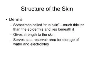
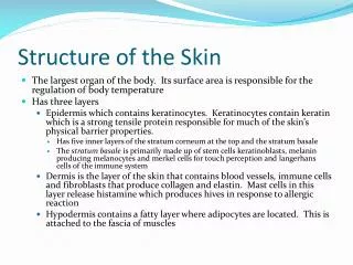
SCOPE The research investigates the behaviour of successive expansions of human skin, in the scalp, lower leg and abdomen. A detailed in vivo analysis is carried out involving five different patients. For each protocol the data of at least four expansions were monitored obtaining at least five measurements relating the volume inserted and pressure inside the skin expander for each expansion. To obtain a constitutive equation that could describe the human skin, several well known constitutive relations were analyzed and Delfino’s constitutive equation was chosen. The skin was considered: incompressible, visco-elastic and homogeneous.
Skin Expansion The first step is the surgery to implant the expander under the skin. Two weeks after the surgery, the process of expansion begins. Weekly a certain volume of saline solution is infiltrated inside the expander. Due to the visco-elastic property of the skin after some time the skin relaxes diminishing the pressure inside the expander.
Pressure Sensor to be coupled to the syringe Numerical Analysis Finite Elements - ABAQUS Newton Raphson Methodology IN VIVO Measurements Five patients; Scalp (2), Lower leg (2) and Abdomen (1).
The process “in vivo” and results To identify the behaviour of the skin due to successive skin expansions it is necessary to measure the pressure inside the skin expander previously, during and after the infiltration of the saline solution inside the expander. (a) (b) (c) Measuring the skin expansion, (a) needle, (b) syringe, (c) pressure sensor.
Patient 1 Step 6 3,0 2,5 Relaxation 2,0 Pressure ( N/cm2) 1,5 1,0 0,5 0,0 0 2 4 6 8 Time (days) Results for Patient 1 (scalp)
Maximum expansion achieved Patient 3 Patient 4 Patient 2 Patient 5
30 25 20 15 10 5 0 0.05 0.1 0.15 0.2 0.25 0.3 0.35 0.4 Results P(N/cm2)*10 Curves relating the maximum percentage of liquid inserted, V**, and the maximum pressure reached at each expansion, blue and green, Scalp ; light blue and red to Lower leg; purple to Abdomen. V** V**=(Vi-Vi-1 )/Vi-1
The ABAQUS Standart finite element mesh was done using linear hybrid membrane elements, (M3D4 e M3D3), with the initial thickness provided by the surgeon and the Newton Raphson Method (MATLAB). Given that the control of the volume inserted inside the skin expander was essential to model the medical procedure, it was made necessary to use fluid finite elements under the membrane, to simulate the infiltration of the skin expander with fluid. Numerical analysis Constitutive Equation: (a e b parameters, I1 first invariant)
Numerical simulation Since the expansions are successive, the final geometry of one expansion, is used as the initial geometry for the next expansion, with zero stress, and the thickness of the modeled skin changes, but not uniformly. Elastic step Relaxation Patient 1 Step 6 3,0 2,5 Relaxation 2,0 1,5 Pressure ( N/cm2) 1,0 0,5 0,0 0 2 4 6 8 Time (days) Step1 In Vivo 3,00 2,50 2,00 1,50 Pressure (N/cm2) 1,00 0,50 0,00 210 220 230 240 250, 260, Volume (ml)
Patient 1 Step 1 3,00 2,50 In vivo Numerical 2,00 Pressure (N/cm2) 1,50 1,00 0,50 0,00 480 500 520 540 560 Volume (ml) Detailed results for patient 1 Expander:rectangular, 13,6cm x 5,5cm x 5,6cm . Mesh:126 quadrilateral linear membrane elements (M3D4) and 126 fluid elements (F3D4). Results of the fitting: in vivo (blue curve) and numerical (pink curve). using Delfino’s exponential function with parameters a = 0.213 MPa and b = 31.5
Volume 380-425ml Volume 110-140ml Volume 80-110ml Volume 425-465ml Volume 465-500ml Finite Elements result for each step Volume 500-538ml
Relaxation (538 ml) Patient 1 Step 6 3,0 2,5 Relaxation 2,0 1,5 Pressure ( N/cm2) 1,0 0,5 0,0 0 2 4 6 8 Time (days) g1=1.0 τ1=1.2 (days)
Parameters for the scalp b = 27.9 + 0.9 e (V*/0.31). a = 1.187V*3 – 1.395 V*2 + 1.075 V* – 0,015.
Parameter a 3,0 2,5 2,0 Scalp a (Mpa) 1,5 Lower Leg Abdomen 1,0 0,5 0,0 0,00 0,50 1,00 1,50 V* Results for the three regions:Parameter a
Parameter b 100,0 90,0 80,0 70,0 Scalp 60,0 Lower Leg 50,0 40,0 Abdomen b 30,0 20,0 10,0 0,0 0,00 0,50 1,00 1,50 V* Results for the three regions:Parameter b
Parameter a Parameter b 0,600 50,0 40,0 0,500 Thickness 0,5cm Thickness 0,5cm 0,400 30,0 Thickness 0,8cm Thickness 0,8cm 0,300 20,0 a MPa b 0,200 10,0 Thickness 1,2cm Thickness 1,2cm 0,100 0,0 0,00 0,50 1,00 1,50 0,000 V* 0,30 0,93 1,09 1,21 V* Parametric study:varying initial thickness Patient3 (lower leg) 0,5cm) Conclusions : Parameter a changes more. It is important to measure precisely the initial thickness of the skin.
Conclusions • The present study is pioneer in its goal to model the human skin in successive skin expansions, obtaining different parameters to characterize the skin as the expansions go on. • Each patient had, between four and six expansion measured, obtaining at least five measurement for each expansion, of each patient. The total data for this research contained more then 100 (volumeXpressure) results. • It is reasonable to understand that as the skin is extended, process done by the use of expanders, the collagen fibers are also extended and in this way here will be a increased resistance for expansion. This can be seen by the increase of parameters a and b , of Delfinos’s constitutive equation, as the expansion goes on.
Conclusions (cont.) • The results, although the number of patients for each region of the body were small, only two, were quite encouraging, and we believe they can be used as an initial guess for the problem. • A further research can provide the type, number and volume of skin expanders necessary to obtain an extra amount of skin to repair a certain medical problem. • Based in the results we can warn the surgeons against to expansion of the skin in regions over elastic foundation, as abdomen or over fatty tissue as on the upper leg.
We are specially grateful to Professor Ivo Pitanguy and his staff who has supported our projects over the years. To the Dr. Henrique Radiwansky for his assistance during the in vivo measurements, in Santa Casa da Misericórdia RJ, (38th Enfermar. - Serviço do Professor I. Pitanguy) and Institute Ivo Pitanguy. Thanks CNPq and FAPERJ for the support with the research projects. Last but not least for the support and enthusiasm of the patients without whom this work was made possible. Acknowledgments