Download
1 / 22
220 likes | 328 Vues
Unification of CytometryML, DICOM and Flow Cytometry Standard. Robert C. Leif *a and Stephanie H. Leif a a XML_Med , a Division of Newport Instruments, 5648 Toyon Road, San Diego, CA 92115 rleif@rleif.com; http:// www.newportinstruments.com Pathology Informatics Boston.

E N D
Unification of CytometryML, DICOM and Flow Cytometry Standard Robert C. Leif*a andStephanie H. Leifa aXML_Med, a Division of Newport Instruments, 5648 Toyon Road, San Diego, CA 92115 rleif@rleif.com; http://www.newportinstruments.com Pathology Informatics Boston
Health Information Technology (HIT) Standards Committee Meeting, Top Three Findings Implementation Starter Kit Hearing Goal is to achieve higher levels of health information exchange by • Creating clear interoperability standards • Disseminating knowledge of increasing availability of tools and utilities • Clarifying the requirements of Meaningful Use for each stage of compliance
Reuse Existing Standards by Translation into XML • DICOM) (http://medical.nema.org/) • Pathology Standard for Digital Microscopy • Supplements (Bruce Beckwith et al.): • 122: Specimen Module and Revised Pathology SOP (Service-Object-Pair) Classes • Specimen, Bar Code, Sampling, Staining • 145: Whole Slide Microscopic Image IOD (Information Object Definition)and SOP Classes • Includes images, Image pyramid storage, Pixel Matrix (x,y,z), focus, Optical Path (incomplete), etc.
Reuse Continued • DICOM Part 18 Web Access to DICOM Persistent Objects (WADO) & its Supplement 148 Web Access to DICOM Persistent Objects”, WADO to Web Services (Out for comment). • Retrieval into XML by generic mechanism for transposing DICOM Header information from binary to XML • Metadata or extract of them based on XPath • XML data types for DICOM Persistent Objects
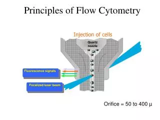
Reuse Continued • Flow Cytometry Standard, FCS 3.1. • New names for the data-types were created and data-types from other CytometryML schemas were reused. • ZIP data file format • Instrument Description
Gating-MLhttp://flowcyt.sourceforge.net/gating/ • Gating (Geometric structures for scene segmentation) • Compensation (Reading values to specific measurements) • Transformations (linear, log, combinations)
Why Extensible Markup Language (XML)? • Since the DICOM and FCS data representations (syntax) are unique, special interface software must be written to interface each to the other and to other software and to hardware. • XML is ubiquitous • XHTML (Web applications) is XML. • A very large amount of data is stored in XML. • XML has been interfaced to virtually all commercial software including databases and can be used in forms and documents.
DICOM Reuse in CytometryMLhttp://www.newportinstruments.com/ • Nomenclature, design, documentation, and data-types. • Tables provide the composition of DICOM data structures (sequences). These become XML complexTypes. • Individual data elements often become XML simpleTypes, which can either be elements and/or attributes.
DICOM Reuse in CytometryML Cont. • The documentation for individual DICOM elements becomes XML documentation elements. • New cytometry data-types are composed in part of DICOM types. • CytometryML consists of 74 schema • Most of which have been validated and are used • 14 XML test documents
Example <annotation><documentation>An identifier of the of the order for the Study. From Table C.7-3GENERAL STUDY MODULE ATTRIBUTES in the draft supplement 122.</documentation></annotation><complexType name="Accession_Number_Type"><simpleContent><extension base="dicom:Short_String_Type"><attribute name="Tag" type="dicom:Tag_Type" use="optional" fixed="00080050"/><attribute name="VR" type="dicom:VR_Type" use="optional" fixed="SH"/></extension></simpleContent></complexType>
DICOM Organization 1 Patient (Medical Record) X Studies Y Series Z Instances The items below the line are the part of the CytometryML schemas that will be discussed.
Optical Path Definition • Positions of excitation optical elements have negative values; • Positions of imaging elements have positive values. • The position of the slide or flow cell that holds the specimen is 0. • The optics go in a positive direction towards the detector and a negative direction towards the light source.
Conclusions • Maximizing reuse including • Designs • Documentation Will • Improve International Medical Informatics • infrastructure and • facilitate interoperability • Increasing safety and minimizing development costs
Conclusions… • It has been possible with XML Schema Definition Language (XSDL) 1.1 to • maximize readability, • create a modular structure, and • strongly typed, reusable data-types.
Conclusions… • Much of FCS & a significant part of DICOM relevant to pathology has been translated into XSDL and then into XML.
Hopes • This infrastructure improvement should benefit the patients while significantly decreasing health care costs. • The combination of the knowledge of pathologists and cytometrists should exceed the sum of its parts.
Future DICOM should be extended in XML and evolve into an XML based standard. • Make XML be a DICOM Coding Scheme