Download
1 / 27
270 likes | 277 Vues
New Perspectives in Molecular Imaging of Cardiovascular Diseases F. Garibaldi - INFN – Roma1. - molecular imaging: the role of radionuclides techniques - building an open, flexible system - cardiovascular diseases ( diagnosis and therapy )

E N D
New Perspectives in Molecular Imaging of Cardiovascular Diseases • F. Garibaldi - INFN – Roma1 • - molecular imaging: the role of radionuclides techniques • - building an open, flexible system • - cardiovascular diseases (diagnosis and therapy) • - detecting vulnerable atherosclerotic plaque • - stem cell therapy of heart infarction • conclusions and outlook
Collaboration Istituto Superiore di Sanita’ E. Cisbani S. Colilli R. Fratoni F. Garibaldi TESA M. Gricia M. Lucentini F. Santavenere S. Torrioli Farmaco G. Marano M. Musumeci Farmaco Ematologia M. Baiocchi Oncologia L. Vitelli INFN – Roma1 F. Cusanno M.L. Magliozzi Jefferson Lab (DOI) S. Majewski D. Weisemberger B. Kross J. Proffit Johns Hopkins University B.Tui Y Wang University of Rome G. De Vincentis
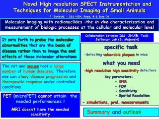
Molecular Imaging • “… the in vivo characterization and measurement of biologic processes at the cellular and molecular level. • - It sets forth to probe the molecular abnormalities that are the basis of diseaserather than to image the end effects of these molecular alterations. • - Imaging of specific molecular targets enables: • earlier detection and characterization of disease; • earlier and direct molecular assessment of treatment effects; • more fundamental understanding of disease processes. • The rat and mouse host a large number of human diseases • Opportunity to study disease progression / therapeutic response • under controlled conditions • non-invasively • in same animal • repetitively Mice advantages: small size, rapid gestation period large litter size, low maintenance costs. Moreover, the mouse genome has been extensively characterized. Gene-targeted "knock-out" and transgenic overexpressionexperiments are performed using mice, rather than rats, but submm spatila resolutin needed!
Ultrasound CT Optical (Bioluminescence, fluorescence) A F Structure A M 0.1 mm Unique !! Doppler Topography µm to mm ~103 cells quantitative Tissue Density, Z A 20-50 µm MRI PET/SPECT F M A F M Radiotracer ~1-2 mm H Concentration <10-12 mole = quantitative 0.1 mm BOLD, DCE Molecular Imaging Modalities -galactocidase 0.1 µmole H / µmole 31P
Patient injected with radioactive drug. Drug localizes according to its metabolic properties. Gamma rays, emitted by radioactive decay, that exit the patient are imaged. Single Photon Detector Module • Collimator • Only gammas that are perpendicular to imaging plane reach the detector • Readout Electronics • Amplify electrical signal and interface to computer • Scintillator • Converts gammas to visible light • Computer decoding procedure • Elaborate signal and gives image output • Photodetector • Convert light to electrical signal
Importance of pixel identification good pixel identification is fundamental for correct digitization affecting spatial resolution and contrast C8 strips M16 (4 x 4) mm2 M64 (2 x 2) mm2
Labr3 Continuoum different performances for different window treatment, diffusing (a), absorbing (b) a b
MCP (Burle) 1.5x1.5 mm2 CsI(Tl) 0.4 -1.0 mm pitch LaBr3
Silicon Drift Detectors Si Pin diode: high QE, simple, economics, but no gain ! noise etc
collimation Compton Camera pet Multipinhole
Performances not good enough for imaging biological process in vivo in small animals (mice) • Small size detectors (high pixellization) • Individual detectors or “perfect” coding Rat Human Required spatial resolution: 1 mm FWHM (1 mm3)) 6 mm FWHM (200 mm3)) 2 mm FWHM (8 mm3) Submillimiter spatial resolution, high sensitivity needed
Cardiovascular diseases • Diagnosys • Detection of cause of occlusiom • Imaging in vivo: detection of vulnerable plaques Infarction • Theraphy • Stem cell theraphy for cardiac repair • Imaging in vivo: monitoring stem cells diffusion, differentiation, grafting, looking at the effects
APPROACH -Transgenic mouse model -APOE-/- mice -Spontaneous growth of atherosclerotic plaques accelerated by fatty diet - Imaging agent • 99mTc-HYNIC-Annexin-V (Binds to apoptotic cells) plaque • ~ 2 mm diameter aorta • ~ 0.5 X 1 X 4 mm3 • Total activity: ~ 1 microCi, • (0.05% of average injected dose of ~2 mCi) 99mTc-Annexin-V SPECT images Excised aorta from 37 weeks mouse Plaque? ApomateTM: Trade name for Hynic Annexin V, North American Scientific, Inc. ApoE-/- mice Photo autoradiography 20 weeks
JHU, Baltimore ISS, Rome
Develop and validate imaging tools for novel cell therapies (e.g. immune cells or stem cels) allowing tracking of cells and assessment of cell fate (e.g. viability, differentiation, migration and therapeutic effects, that could be tested in animal models with a view towards translational medicine. The development tools should provide specific and quantifiable information with respect, for example, to cell homing, functional read-outs, tracking of differentiation or immune systemresponse .FP7 These reports underscore the need for a greater understanding of the mechanisms underlying stem cell biology and cellular reparative therapy, and their potential uses in the post-infarction state.
- Optical 10e15 to 10e17 mol/l - PET,SPECT 10e11 to 10e12 mol/l - MRI 10e5 mol/l direct labeling: labels may be diluted upon cell division, making these cells invisible; and labels may efflux from cells or may be degraded over time. alternative approach: stable transfection of cells with a reporter gene, such as herpes simplex virus type-1thymidine kinase (HSV1-tk), whose expression can be visualized using a radioactive PET or SPECT reporter probe (phosfphorilates --> TK --> triphosphate --> cells) PET and SPECT imaging can be used to assess cell trafficking, function, and efficacy, using methods which are easily translatable to humans. Reporter gene approaches are particularly valuable, as they provide information not only on cell trafficking, but also on cellular function and survival
Dual labeling - Optical imaging techniques provide high spatial resolution and permit tracking of stem cells but are limited to preclinical use - Magnetic resonance imaging methods permit good spatial resolution but limited detectability - Nuclear techniques, including reporter genes and direct cellular radiolabeling, afford very good detectability but more limited spatial resolution A multimodality approach using combined PET or SPECT and MRI agents may ultimately prove most useful in clinical settings. P. Acton and al.
and Multimodality (Zhou, Acton) SPECT/MRI OPTICAL/PET (Gambir)
Detector ~ 100x100 mm2 • Intrinsic reaolution: 1.2 - 1.5 mm • pinhole (0.5 mm) rhigh resolution : ~ 0.8 mm • M= 3 ==> FoV ~ 33 x 33 mm2 • Perfusion Imaging • Tracking and homing of stem 8 cm 2.5 cm wall thickness ~ 0.8 mm !!!! mouse: C57 BL/6, male age: ~ 12 weeks weight: ~31.5 g stem cells:~ 6*104 murine (SCA+/KIT+ Tc99m-HMPAO (28 microCi)
Reconstruction Images of Mouse Perfusion Scan (I) Trans-axial Axial Trans-axial Axial OS-EM, 6 subsets, 2 iterations, post-smoothed by Butterworth filter (cutoff=0.12, order=8), voxel size = 0.25 mm, image dimension 90x90. Reconstruction Images of Mouse Perfusion Scan (II)
Tail vein injection Peritoneum injection • It is extremely difficult if not impossible to use the tail vein for radiotracer many times. An alternative route of delivery is needed, but, how much of will arrive to heart? Let’s look at the peritoneum. • It works, but there is a price to pay, the uptake is decreased (a factor of ~ 2). • We have to maximize the efficiency • More detectors • Multipinhole
Conclusions Importance of molecular imaging in the biomedical research panorama: crucial role of radionuclides techniques Multidisciplinary approach mandatory Multimodality (“new” photosensors (SiPm?)) • atherosclerosys: • looking for smaller plaques “earlier” detection (other mechnisms, other radiotracers) - stem cells: - selecting “right” cells - monitoring diffusion, differentiation, grafting etc - looking at the effects ===> multimodality (optical, SPECT, MRI, )
Outlook • the challenge:120 pixel/100 mm, 8 modules 150 X 100 mm2 • ---> FOV 50 x 33 (M=3))
12C(e,e’K)12BL Aerogel Kaon selection RICH Kaon selection Freon/CsI RICH detector (like ALICE) Spectroscopy analysis of 12B: Aerogel vs. RICH K-selection Hermes areogel RICH
Important parameters fordetectability/visibility scintillator electronics, DAQ efficiency collimation time (and modality) uptake (radiopharmacy) spatial resolution detector intrinsic properties modality (compression) pixel dim/n.of pixels . Uniformity of p.h.response (affecs the overall en res. and the energy window seection) CsI(Na) CsI(Tl) Bialkali PMT Bialkali PMT fotofraction