Download

1 / 7
70 likes | 82 Vues
M. Bulent Balioglu, Sebahat Aydil, Pınar Benek , Akif Albayrak, Yunus Atici, Y. Emre Akman , Deniz Kargın , M. Temel Tacal, M. Akif Kaygusuz. Gait Analysis in Patients With Congenital Scoliosis.

E N D
M. Bulent Balioglu, SebahatAydil, Pınar Benek, Akif Albayrak, Yunus Atici, Y. EmreAkman, Deniz Kargın, M. Temel Tacal, M. Akif Kaygusuz Gait Analysis in Patients With Congenital Scoliosis BaltalimanI Metin sabancI Disease of the Bone Education and Research Hospital İstanbul, Turkey
Author Disclosure Information • M.B. Balioglu None • S. Aydil None • P. Benek None • A. Albayrak None • Y. Atici None • Y.E. Akman None • D. Kargın None • M.T. Tacal None • M.A. Kaygusuz None
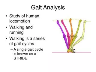
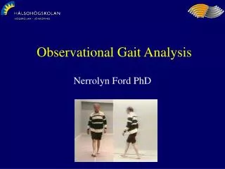
Introduction • We planed to described gait abnormalities in congenital scoliotic patients. • The goal of this study is to describe gait abnormalities in congenital scoliotic patients with 3D computed gait analysis system.
Methods • We assessed the gait pattern of 12 patients with congenital scoliosis(5 M, 7 F). • The mean age was 9.8 years (5-14). • A three-dimensional motion capture system was used to track a full body marker set that was placed on the participant’s body. • The 3D-segment positions of the head, trunk, pelvis, individual joint angles of the lower extremities, and spatial-temporal parameters were computed during walking. • We compared the kinematic graphs and spatial-temporal parameters of congenital scoliotic patients with the healthy subjects at the same age group.
Results • All patients walked at a normal velocity (median: 1.06 ± 0.16 m/s) and the timing of the individual gait phases was normal and symmetrical for the whole group. • Only difference in spatial-temporal parameters was cadence (median: 122 ± 15.0 steps/min) and greater than healthy subjects (median: 117 ± 13.7 steps/min). • Sagittal plane pelvis, knee and ankle motion followed a physiological pattern although ankle motion showed wide range during the gait cycle. • The sagittal plane of hip motion mean trace was observed in a flexed pattern during the whole gait cycle.
Results • The hip motion in the coronal plane was observed in an abducted pattern while pelvis, knee and ankle motion were within normal range. • Transvers plane kinematics of the pelvis and lower extremities were within normal range. • The transvers and coronal plane kinematics of head, neck and trunk motion’s mean traces were within the normal range although graphs were observed in a wide range. • The sagittal plane kinematics graphs of head, neck and trunk were different from the healthy subjects.
Conclusions • The most significant differences were seen in the sagittal and coronal plane of hip, and the sagittal plane of head, neck and trunk. • All other graphs were observed within normal range in the kinematics of the congenital scoliotic patients. • Further studies are needed to identify the gait patterns in this patient group.