Download
1 / 50
510 likes | 526 Vues
Lodish • Berk • Kaiser • Krieger • Scott • Bretscher •Ploegh • Matsudaira. MOLECULAR CELL BIOLOGY SIXTH EDITION CHAPTER 13 Moving Proteins into Membranes and Organelles.

E N D
Lodish • Berk • Kaiser • Krieger • Scott • Bretscher •Ploegh • Matsudaira MOLECULAR CELL BIOLOGY SIXTH EDITION CHAPTER 13 Moving Proteins into Membranes and Organelles
A. How a given protein could be targeted to only specific membrane?B. How relatively large protein molecules could be translocated across a membrane without disrupting the bilayer?
Answer questions regarding targeting1. What is the nature of the signal sequence?2. What is the receptor for the signal sequence?3. What is the structure of the translocation channel?4. What is the source of energy?
13.1 Translocation of Secretory proteins across the ER membraneextracellular spacelysosomal lumenER, Golgi lumen (soluble proteins)Pancreatic acinar cell
Labeling experiments demonstrated that secretory proteins are localized to the ER shortly after synthesis
A hydrophobic N-terminal signal sequence targets nascent secretory protein to the ERa. 16-30 residue ER signal sequence(SS) (contain 16-30 sequence residue followed by hydrophobic aa)b. only present in nascent protein not in mature proteinc. mutation in SS, the protein stays in cytosol
2. Cotranslational translocation is initiated by two GTP-hydrolyzing proteinsa. SRP: cytosolic ribonuclear protein binds to signal sequence to the large ribosomal subunit and to SRP receptorb. SRP receptor is in the ER membrane.
SRP can inhibit the elongation of nascent chain until it binds to its receptor • Then the SRP release, the polypaptide starts to elongate
3.Passage of growing polypeptides through the translocon is driven by energy released during translationco-translational transport
4. ATP hydrolysis powers post-translational translocation of some secretory proteins in yeast
13.2 Insertion of proteins into the ER membrane (orientation preserved)1. Several topological classes of integral membrane proteins are synthesized on the ER.Topology: a. span how many times and the orientation of these membrane-span segments (membrane span segment itself:20-25hydrophobic aa) b . Internal stop-transfer and signal-anchor sequence determine topology of single- pass proteins.2. covalently bind to phospholipid
2. Internal stop-transfer and signal-anchor sequence determine topology of single-pass proteins.
Type IV: Multipass proteins have multiple internal topogenic sequences
2. A phospholipid anchor tethers some cell-surface proteins to the membrane.By Glycosylphosphatidylinositol (GPI): an amphipathic moleculeGPI is synthesized and attached to the ER membrane (type I)
5. The topology of a membrane protein often can be deduced from sequenceEach segment of proteins → hydrophilic environment∆G : -, means hydrophilic sequence computer program generate hydropathy profiles
13.3 Protein modifications, folding, and quality control in the ER
1. Addition and processing of CHO (glycolyzation) in the ER and Golgi (ER mainly)2. Formation of disulfide bonds in the ER(only in ER)3. Proper folding of polypeptide chains and multisubunit proteins in the ER (only in ER) 4. Specific proteolytic cleavages in the ER, Golgi, and secretory vesicles.
1. A preformed N-linked oligosaccharides is added to many proteins in the RER • In plant, animal, and signal-celled eukaryotes have the similar precusors: 3 Glc, 9 mannose, 2 GlcNAc
2. Oligosaccharide side chains may promote folding and stability of glycoproteins a. glycosylated fibronectin is not digested easilyb. CAM extensively glycosylated increasing the adherence of the blood cellsc. recognition (ABO blood type)d. if hemagglutinin precusor polypeptide (HA0) can not fold properly, can not form trimer, and only can stay in the ER
3. Disulfide bonds are formed andrearranged by proteins in the ER lumen.S-S proteins only found in secretory proteins(in ER luman)protein disulfide isomerase (PDI) : rich in liver and pancreas
4. Chaperons and other ER proteins facilitate folding and assembly of proteins.In vitro: protein folding, several hrsin vivo: only few mins
Bip:prevent misfolding or forming aggregatePD1(protein disulfide isomerase): proper folding by stabilizing S-S bondLectin: carbohydrate-binding proteins (calnexin, calreticulin) bind to nascent chain in ER (HA assembly)Peptidyl-prolyl isomerases : accelerate the rotation about peptidyl-prolyl bonds in unfolded segment and accelerate folding.
Folding and assembly of hemagglutinin (HA0) trimer in the ERHA is made by host cell and form the spike form on the surface of influenza virusHA0is formedin the ER, and then form trimer (HA0, HA0, HA0). Then each HA0 cleavage to 2 polypeptide chains HA1, HA2 and form three copies of HA1, HA2 in Golgi Fig. 13-20
5. Improperly folded proteins in the ER induce expression of protein-folding catalystsBip, calnexin (folding catalysts)a. assisting in the folding of normal proteins by preventing aggregationb. binding to irreversible misfolded proteinsUnfolded-protein response: Ire-1 protein
α1- anti-trypsin produced from hepatocytes and macrophages and inhibit trypsin and blood protease, elastase. If α1- anti-trypsin has point mutation, stored in the ER, can not inhibit trypsin and elastase → elastase can digest lung tissue → emphysema (genetic disease)
6. Unassembled or misfolded proteins in the ER are often transported to the cytosol for degradation (ubiquitin-mediated proteolytic pathway). ubiquitinylating enzymes localized to the cytosolic faceof the ER add ubiquitin to misfolded ER proteins as they exit the ER. (need ATP) (fig. 3.29 and chapter 14)
2004 nobel prize • Aaron Ciechanover, Avram Hershko, I Irwin Rose
Export of bacterial proteins1. Cytosolic SecA ATPase pushes bacterial polypeptides (toxins or pili) through translocons into the periplasmic space.2. All bacterial proteins that are translocated across the inner membrane soon after their synthesis in the cytosol is complete but before they are folded into their final formation
3. Pathogenic bacteria can inject proteins into animal cells via Type III secretion.Yersinic pestis (Plague): destroy macrophage of human being. Apparatus inject small set of proteins into macrophage
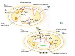
13.4 Sorting of proteins to mitochondria and chloroplasts layers organelles: DNA, RNA, ribosome, proteins.
1. Amphipathic N-terminal signal sequence direct proteins to the mitochondrial matrixmatrix-targeting sequence (20-50 aa hydrophobic acid, and some + or OH aa)
2. Mitochondrial protein import requiresouter-membrane receptors and translocons in both membranes.
3. Studies with chimeric proteins demonstrate important features of mitochondria import.Use recombinant DNA to form chimeric proteinsUnfolded proteinsMatrix target sequence
4. Multiple signals and pathways target proteins to submitochondrial compartments.
5. Targeting of chloroplast stromal proteins is similar to import of mitochondrial matrix proteins.a. Calvin cycle (rubisco L subunit made in stroma, others made from nucleus) b. N-terminalstromal import sequence
13.5 Sorting of peroxisomal proteins (lack DNA)1. Cytosolic receptor targets proteins with a SKL(serine, lysine, leucine) sequence at the C-terminus into the peroxisomal matrix.2. Peroxisomal membrane and matrix proteins are incorporated by different pathways.
Peroxisomal membrane and matrix proteins are incorporated by different pathways
13.6 transport into and out of the nucleus • Nuclear pores • Nuclear localization sequences (NLSs) • In a folded state different from ER transport • Proteins, ribonuclear proteins (ribosomes) • mRNA • Mechanisms of import and export are different
1. Large and small molecules enter and leave the nucleus via nuclear pore complexes • Nuclear pore complex (NPC) • 60-80 million Da (16 times large than ribosome) • Multiple copies of some 30 different proteins, nucleoporins • Ions, small molecules, and globular proteins (20-40kD) can passively diffuse through central aqueous region • Large macromolecules are actively transported through the NPCwith thr assistance of soluble transport proteins that bind macromolecules and also interact with nucleoporins.
Small molecules (20-40 kDa): passive diffusion • Large proteins and ribonucleoproteins actively transported through pores with assistance of soluble transporter proteins
2. Importins transport proteins containing nuclear-localization signals into the nucleus • Histones, TF, DNA polymerases, RNA polymerases • NLS • Simian virus 40 (SV 40) mutants: large T-antigen accumulates in the cytoplasm • 7 basic aa at c-terminus • Engineered 7 aa to a cytoplasmic protein demonstrated it directs transport into nucleus • Other NLSs were found
Nuclear-localization signal (NLS) directs proteins to the cell nucleas
Importins • αsubunit: binds to a basic NLS in a cargo protein to be transported into the nucleus • Βsubunit: interacts with proteins in the nuclear pore to shuttle the cargo protein through it
3. Exportins transport proteins containing nuclear-export signals out of the nuclear3 • Nuclear-export signal (NES) (vs NLS) • Engineered hybrid genes encoding a restricted nuclear protein fused to various segments of a protein that shuttles in and out of the nucleus • Identified at least three classes of NESs a. a leucine-rich sequence found in PKI and in the Rev of HIV virus b. two hnRNPs • The functionally significant structural features that specify nuclear export remain poorly understood.
Leucine-rich NES • Exportin (nuclear-export receptor) in nucleus (exportin-1) binds to Ran.GTP and then binds to NES in a cargo protein