Download
1 / 19
200 likes | 428 Vues
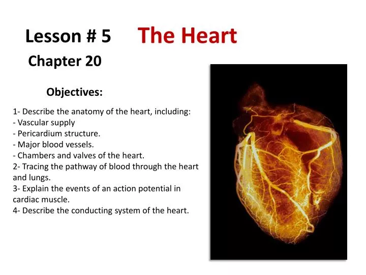
The Heart. Lesson # 5. Chapter 20. Objectives:. 1- Describe the anatomy of the heart, including: - Vascular supply Pericardium structure. Major blood vessels. - Chambers and valves of the heart.

E N D
The Heart Lesson # 5 Chapter 20 Objectives: • 1- Describe the anatomy of the heart, including: • - Vascular supply • Pericardium structure. • Major blood vessels. • - Chambers and valves of the heart. • 2- Tracing the pathway of blood through the heart and lungs.3- Explain the events of an action potential in cardiac muscle. • 4- Describe the conducting system of the heart.
An Introduction to the Cardiovascular System Systemic Circuit It supplies blood to every organ of the body, including the heart itself. Cardiovascular System Pulmonary Circuit Aorta Pulmonary Pulmonary Vena cava It carries blood to the lungs for gas exchange and returns it to the heart. Arteries (2) Veins (4) Blood Vessels Arteries They carry blood away from the heart. Veins They carry blood back to (toward) the heart. Capillaries They connect the arteries with the veins.
Pulmonary Circuit Gas exchange It carries blood to the lungs for gas exchange and returns it to the heart. O2rich blood through VEINS O2poor blood through ARTERIES Systemic Circuit It supplies blood to every organ of the body, including the heart itself. O2poor blood through VEINS O2rich blood through ARTERIES
GasExchange Systemic Pulmonary Circuit Circuit Capillary Lung Venule Arteriole Pulmonary arteries Pulmonary veins O2 poor, CO2 rich blood O2 rich, CO2 poor blood Wastes Nutrients O2 O2 CO2 CO2 Venae cavae Aorta Capillary Tissue Venule Arteriole
Cardiovascular System: Pulmonary Circuit It carries blood to the lungs for gas exchange and returns it to the heart Systemic Circuit It supplies blood to every organ of the body, including the lungs and the heart itself Blood Vessels: Arteries They carry blood away from the heart Veins They carry blood back to (toward) the heart Capillaries They connect the arteries with the veins
The heart is located in the chest cavity, surrounded by the pericardial sac, in the anterior portion of the mediastinum.
The Pericardium Pericardial cavity The pericardium is a double-walled sac (pericardial sac) that encloses the heart. Parietal pericardium Visceral pericardium (epicardium) Pericarditis is a disorder caused by inflammation of the pericardium, the sac-like covering of the heart. Mesothelium Areolar tissue Fibrous tissue Pericarditis can be caused by bacterial, fungal, or viral infections. It may also be a result of injury or trauma to the chest, esophagus, or heart. Pain occurs as a result of the inflamed pericardium rubbing against the heart.
Superficial Anatomy of the Heart Right pulmonary veins Left pulmonary veins Pulmonary trunk Superior vena cava Lungs Superior vena cava Coronary sulcus Aorta Left atrium Coronary sinus Anterior interventricular sulcus Left atrium Right atrium Right atrium Left ventricle Inferior vena cava Right ventricle Inferior vena cava Left ventricle Right ventricle Coronary sulcus Coronary sinus Posterior interventricular sulcus Anterior surface Posterior surface
The Heart Wall Parietal pericardium Areolar tissue Areolar tissue Pericardial cavity MYOCARDIUM (cardiac muscle tissue) ENDOCARDIUM EPICARDIUM Endothelium Mesothelium Visceral pericardium Mesothelium Areolar tissue Fibrous tissue Endocarditis is inflammation of the inside lining of the heart chambers and heart valves (endocardium). Most people who develop endocarditis have heart disease of the valves.
Internal Anatomy and Organization Gas exchange Superior vena cava Poor oxygen blood Pulmonary arteries Coronary sinus Reach oxygen blood Inferior vena cava Pulmonary veins (4) RIGHT ATRIUM LEFT ATRIUM Aorta RIGHT VENTRICLE LEFT VENTRICLE To the rest of the body
Superior vena cava It drains oxygen-poor blood from tissues and organs superior to the diaphragm to the right atrium. Aorta Pulmonary trunk It carries oxygen-rich blood from the left ventricle to the whole body. It carries oxygen-poor blood from the right ventricle to the lungs. Pulmonary veins (4) Inferior vena cava They carry oxygen-rich blood from the lungs to the left atrium. It drains oxygen-poor blood from tissues and organs inferior to the diaphragm to the right atrium. Coronary sinus (no shown) It drains oxygen-poor blood from the heart tissues to the right atrium.
The Heart Valves The heart has two pairs of one-way valves that prevent the backflow when the chambers contract Pressure Pressure It prevents back flow of blood from the LV to the LA It prevents back flow of blood from the RV to the RA Left AV (bicuspid) valve Right AV (tricuspid) valve
The Heart Valves The heart has two pairs of one-way valves that prevent the backflow when the chambers contract It prevents back flow of blood from the pulmonary trunk to the RV Aortic semilunar valve Pressure Pressure Pulmonary semilunar valve It prevents back flow of blood from the aorta to the LV
Ligamentum arteriosum Four openings of the pulmonary veins Remnant of ductus arteriosum Opening of superior vena cava Aortic arch (Remnant of foramen oval) Pulmonary trunk Fossaovalis Pulmonary veins Opening of coronary sinus Aortic semilunar valve Opening of inferior vena cava It prevents back flow of blood from the aorta to the LV Left AV valve or bicuspid valve Right AV valve or tricuspid valve It prevents back flow of blood from the RV to the RA (mitral valve) It prevents back flow of blood from the LV to the LA Pulmonary semilunar valves It prevents back flow of blood from the pulmonary trunk to the RV
Cusps Chordae tendineae Papillary muscle Trabeculae carneae Transverse section, superior view
Circumflex artery Left coronary artery Anterior I-V artery Posterior I-V artery Right coronary artery Right coronary artery Marginal arteries Left coronary artery Coronary sinus Great cardiac vein Circumflex artery Anterior interventricular artery Posterior interventricular artery Small cardiac vein Marginal artery Middle cardiac vein The Blood Supply to the Heart Coronary Arteries and Coronary Veins
Atrioventricular bundle or bundle of His Right and left bundle branches Sinoatrial node (SA node) Atrioventricular node (AV node) Purkinje fibers The Conducting System It connects electrically the atria to the ventricles. They conduct the impulse to the Purkinje fibers. It establishes the heart rate (pacemaker). It delays the impulses to allow the atria to finish contracting before the ventricles start to contract. They conduct the impulse to the lateral walls of the ventricles allowing the contraction to spread from the apex to the base.
Impulse Conduction through the Heart SA node fires and atrial activation begin. Time 0 5 2 3 4 5 5 1 2 3 4 1 Stimulus spreads across the atrial surfaces and reaches the AV node. Elapsed time: 50 msec There is a 100 msec delay at the AV node. Atrial contraction begins. AV node fires. Elapsed time : 150 msec The impulse travels along the inter-ventricular septum within the AV bundle and the bundle branches to the Purkinje fibers. Elapsed time: 175 msec The impulse is distributed by Purkinje fibers and relayed through the ventricular myocardium. Atrial contraction is completed and ventricular contraction begins. Elapsed time: 225 msec