Download
1 / 59
590 likes | 722 Vues
Animal Reproduction & Development (Ch. 46, 47). A “homunculus” inside the head of a human sperm. Sexual & asexual reproduction. Asexual offspring all have same genes (clones) no variation Sexual gametes (sperm & egg) fertilization mixing of genes variation. Parthenogenesis.

E N D
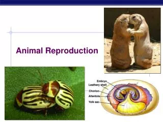
Animal Reproduction & Development (Ch. 46, 47)
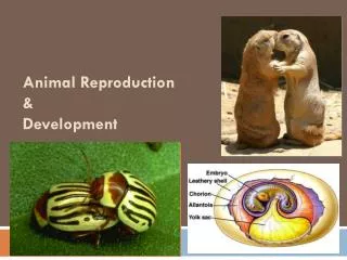
Sexual & asexual reproduction • Asexual • offspring all have same genes (clones) • no variation • Sexual • gametes (sperm & egg) fertilization • mixing of genes variation
Parthenogenesis • Development of an unfertilized egg • honey bees • drones = males produced through parthenogenesis haploid • workers & queens = females produced from fertilized eggs diploid queen worker drone
Honey bee eggs hatch regardless of whether the are fertilized. The female bees--queens & workers--develop from fertilized eggs that contain 32 chromosomes. These 32 chromosomes consist of two sets of 16, one set from each parent. Hence female bees are said to be diploid in origin. The males (drones) develop from unfertilized egg which contain only one set of 16 chromosomes from their mother. Drones are thus haploid in origin This reproduction by the development of unfertilized eggs is called parthenogenesis • Drones develop by parthenogenesis from unfertilized eggs that the queen produces by withholding sperm from the eggs laid in large drone cells. Drones lack stings and the structures needed for pollen collection; in the autumn they are ejected by the colony to starve, unless the colony is queenless. New drones are produced in the spring for mating. • Both queens and workers are produced from fertilized eggs. Queen larvae are reared in special peanut-shaped cells and fed more of the pharyngeal gland secretions of the nurse bees (bee milk or royal jelly) than the worker larvae are. The precise mechanism for this caste differentiation is still uncertain. Although workers are similar in appearance and behavior to other female bees, they lack the structures for mating. When no queen is present to inhibit the development of their ovaries, however, workers eventually begin to lay eggs that develop into drones.
Different strokes… gay penguins parthenogenesis in aphids “lesbian” lizards sex-change in fish
Hermaphrodites • Having functional reproductive system of both sexes earthworms mating flat worm
Fertilization • Joining of egg & sperm • external • usually aquatic animals • internal • usually land animals
Development • External • development in eggs • fish & amphibians in water • soft eggs= exchange across membrane • birds & reptiles on land • hard-shell amniotic eggs • structures for exchange of food, O2 & waste • sharks & some snakes • live births from eggs • Internal • placenta • exchange food & waste • live birth
Adaptive advantages? • What is the adaptive value of each type of sexual reproduction • number of eggs? • level of parental of care • habitat?
Reproductive hormones • Testosterone • from testes • functions • sperm production • 2° sexual characteristics • Estrogen • from ovaries • functions • egg production • prepare uterus for fertilized egg • 2° sexual characteristics LH &FSH testesorovaries
Male reproductive system • Sperm production • over 100 million produced per day! • ~2.5 million released per drop!
Spermatogenesis Testis Epididymis Germ cell (diploid) Coiled seminiferous tubules 1° spermatocyte (diploid) MEIOSIS I 2° spermatocytes (haploid) MEIOSIS II Vas deferens Spermatids (haploid) Spermatozoa Cross-section of seminiferous tubule
Menstrual cycle LH FSH Hypothalamus egg development ovulation = egg release GnRH corpus luteum Pituitary FSH & LH estrogen progesterone Ovaries lining of uterus estrogen Body cells days 0 7 14 21 28
Egg maturation in ovary • Corpus luteum • produces progesterone to maintain uterine lining
Female hormones • FSH & LH • release from pituitary • stimulates egg development & hormone release • peak release = release of egg (ovulation) • Estrogen • released from ovary cells around developing egg • stimulates growth of lining of uterus • lowered levels = menstruation • Progesterone • released from “corpus luteum” in ovaries • cells that used to take care of developing egg • stimulates blood supply to lining of uterus • lowered levels = menstruation
Oogenesis What is theadvantage ofthis development system? • Unequal meiotic divisions • unequal distribution of cytoplasm • 1 egg • 2 polar bodies Meiosis 1 completed during egg maturation ovulation Meiosis 2 completed triggered by fertilization Put all your eggin one basket!
Fertilization • fertilization • cleavage • gastrulation • neurulation • organogenesis
Fertilization • Joining of sperm & egg • sperm head (nucleus) enters egg
What is the effect of sperm binding on Ca2+ distribution in the egg? EXPERIMENT A fluorescent dye that glows when it binds free Ca2+ was injected into unfertilized sea urchin eggs. After sea urchin sperm were added, researchers observed the eggs in a fluorescence microscope. RESULTS 10 sec after fertilization 1 sec before fertilization 30 sec 20 sec Spreading wave of calcium ions Point of Sperm entry The release of Ca2+ from the endoplasmic reticulum into the cytosol at the site of sperm entry triggers the release of more and more Ca2+ in a wave that spreads to the other side of the cell. The entire process takes about 30 seconds. CONCLUSION 500 m
Timeline for the fertilization of sea urchin eggs Binding of sperm to egg 1 2 Acrosomal reaction: plasma membrane depolarization (fast block to polyspermy) 3 4 6 Seconds 8 Increased intracellular calcium level 10 20 Cortical reaction begins (slow block to polyspermy) 30 40 50 Formation of fertilization envelope complete 1 2 Increased intracellular pH 3 4 5 Increased protein synthesis Minutes 10 Fusion of egg and sperm nuclei complete 20 30 Onset of DNA synthesis 40 60 First cell division 90
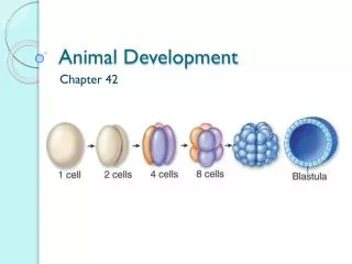
Cleavage • Repeated mitotic divisions of zygote • 1st step to becoming multicellular • unequal divisions establishes body plan • different cells receive different portions of egg cytoplasm & therefore different regulatory signals
Cleavage • zygote morula blastula • establishes future development zygote gastrulation blastula morula
Gastrulation gastrulation inprimitive chordates • Establish 3 cell layers • ectoderm • outer body tissues • skin, nails, teeth,nerves, eyes, lining of mouth • mesoderm • middle tissues • blood & lymph, bone & notochord, muscle, excretory & reproductive systems • endoderm • inner lining • digestive system, lining of respiratory, excretory & reproductive systems ectoderm mesoderm endoderm protostome vs. deuterostome
Testing… All of the following correctly describe the fate of the embryonic layers of a vertebrate EXCEPT A. neural tube and epidermis develop from ectoderm B. linings of digestive organs and lungs develop from endoderm C. notochord and kidneys develop from endoderm D. skeletal muscles and heart develop from mesoderm E. reproductive organs and blood vessels develop from mesoderm
Testing… In a study of the development of frogs, groups of cells in the germ layers of several embryos in the early gastrula stage were stained with five different dyes that do not harm living tissue. After organogenesis (organ formation), the location of the dyes was noted, as shown in the table below. Tissue Stain Brain Red Notochord Yellow Liver Green Lens of the eye Blue Lining of the digestive tract Purple
Neurulation • Formation of notochord & neural tube • develop into nervous system develops into CNS (brain & spinal cord) Neural tube Notochord develops intovertebral column
Organogenesis Umbilical blood vessels Mammalian embryo Chorion Bird embryo Amnion Yolk sac Allantois Fetal blood vessels Placenta Maternal blood vessels
Four stages in early embryonic development of a human Endometrium (uterine lining) Inner cell mass Trophoblast Blastocoel Blastocyst reaches uterus. 2 1 3 4 Expanding region of trophoblast Maternal blood vessel Epiblast Hypoblast Trophoblast Blastocyst implants. Expanding region of trophoblast Amniotic cavity Amnion Epiblast Hypoblast Chorion (from trophoblast) Extraembryonic membranes start to form and gastrulation begins. Extraembryonic mesoderm cells (from epiblast) Yolk sac (from hypoblast) Amnion Allantois Chorion Ectoderm Mesoderm Endoderm Yolk sac Gastrulation has produced a three- layered embryo with four extraembryonic membranes. Extraembryonic mesoderm
Sources of developmental information for the early embryo Unfertilized egg cell Sperm Molecules of another cyto- plasmic deter- minant Molecules of a a cytoplasmic determinant Fertilization Zygote (fertilized egg) Mitotic cell division Two-celled embryo (a) Cytoplasmic determinants in the egg. The unfertilized egg cell has molecules in its cytoplasm, encoded by the mother’s genes, that influence development. Many of these cytoplasmic determinants, like the two shown here, are unevenly distributed in the egg. After fertilization and mitotic division, the cell nuclei of the embryo are exposed to different sets of cytoplasmic determinants and, as a result, express different genes. Nucleus
Early embryo (32 cells) Signal transduction pathway NUCLEUS Signal receptor Signal molecule (inducer) (b) Induction by nearby cells. The cells at the bottom of the early embryo depicted here are releasing chemicals that signal nearby cells to change their gene expression.
Cell signaling and induction during development of the nematode Epidermis 2 Posterior Anterior Signal protein 1 4 Gonad Anchor cell Receptor 3 Signal protein EMBRYO 4 3 Vulval precursor cells Signal Anterior daughter cell of 3 Posterior daughter cell of 3 Inner vulva Outer vulva ADULT Will go on to form muscle and gonads Will go on to form adult intestine Epidermis (a) (b) Induction of the intestinal precursor cell at the four-cell stage. Induction of vulval cell types during larval development.
The effect of the bicoid gene, a maternal effect (egg-polarity) gene in Drosophila Tail Head T1 A8 T2 A7 T3 A6 A1 A5 A2 A4 A3 Wild-type larva Tail Tail A8 A8 A7 A6 A7 Mutant larva (bicoid) (a) Drosophila larvae with wild-type and bicoid mutant phenotypes. A mutation in the mother’s bicoid gene leads to tail structures at both ends (bottom larva). The numbers refer to the thoracic and abdominal segments that are present.
Egg cell Nurse cells Developing egg cell 1 bicoid mRNA Bicoid mRNA in mature unfertilized egg 2 Fertilization 100 µm Translation of bicoid mRNA Bicoid protein in early embryo 3 Anterior end (b) Gradients of bicoid mRNA and bicoid protein in normal egg and early embryo.
Conservation of homeotic genes in a fruit fly and a mouse Adult fruit fly Fruit fly embryo (10 hours) Fly chromosome Mouse chromosomes Mouse embryo (12 days) Adult mouse
Effect of differences in Hox gene expression during development in crustaceans and insects Genital segments Abdomen Thorax Thorax Abdomen
Vertebrate limb development Anterior (a) Organizer regions. Vertebrate limbs develop from protrusions called limb buds, each consisting of mesoderm cells covered by a layer of ectoderm. Two regions, termed the apical ectodermal ridge (AER, shown in this SEM) and the zone of polarizing activity (ZPA), play key organizer roles in limb pattern formation. AER ZPA Limb bud Posterior Apical ectodermal ridge 50 µm (b) Wing of chick embryo. As the bud develops into a limb, a specific pattern of tissues emerges. In the chick wing, for example, the three digits are always present in the arrangement shown here. Pattern formation requires each embryonic cell to receive some kind of positional information indicating location along the three axes of the limb. The AER and ZPA secrete molecules that help provide this information. Digits Anterior Ventral Proximal Distal Dorsal Posterior
What role does the zone of polarizing activity (ZPA) play in limb pattern formation in vertebrates? EXPERIMENT ZPA tissue from a donor chick embryo was transplanted under the ectoderm in the anterior margin of a recipient chick limb bud. Anterior New ZPA Donor limb bud Host limb bud ZPA Posterior RESULTS In the grafted host limb bud, extra digits developed from host tissue in a mirror-image arrangement to the normal digits, which also formed (see Figure 47.26b for a diagram of a normal chick wing). CONCLUSION The mirror-image duplication observed in this experiment suggests that ZPA cells secrete a signal that diffuses from its source and conveys positional information indicating “posterior.” As the distance from the ZPA increases, the signal concentration decreases and hence more anterior digits develop.
Sex determination Zygote Sperm Develop in early embryo Y Testes XY Ovum X SRY Seminiferous tubules Indifferent gonads Leydig cells No SRY X Ovaries Ovum XX (Follicles do not develop until third trimester) X Sperm Zygote
Placenta • Materials exchange across membranes
Placental circulation Maternal veins Maternal arteries Placenta Maternal portion of placenta Umbilical cord Chorionic villus containing fetal capillaries Fetal portion of placenta (chorion) Maternal blood pools Uterus Umbilical arteries Fetal arteriole Umbilical vein Fetal venule Umbilical cord
Human fetal development 4 weeks 7 weeks
Human fetal development 10 weeks
Human fetal development 12 weeks 20 weeks
Human fetal development • The fetus just spends much of the 2nd & 3rd trimesters just growing …and doing various flip-turns & kicks inside amniotic fluid Week 20
Human fetal development • 24 weeks (6 months; 2nd trimester) fetus is covered with fine, downy hair called lanugo. Its skin is protected by a waxy material called vernix
Human fetal development • 30 weeks (7.5 months) umbilical cord