Download
1 / 1
10 likes | 165 Vues
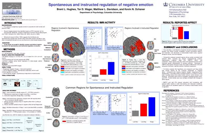
Columbia Psychology SCAN Unit. http://www.scan.psych.columbia.edu/. 5. r = 0.36. 6. r = 0.44. 5. 4. 3. 0. Reapp Neg > Look Neg BOLD. 2. IFG. 1. 0. VMPFC. -1. -5. Hippocampus. 1. 1. 1.5. 0. 0.5. 2. 1.5. 2. 2.5. 3. Drop in Negative Affect

E N D
Columbia Psychology SCAN Unit http://www.scan.psych.columbia.edu/ 5 r = 0.36 6 r = 0.44 5 4 3 0 Reapp Neg > Look Neg BOLD 2 IFG 1 0 VMPFC -1 -5 Hippocampus 1 1 1.5 0 0.5 2 1.5 2 2.5 3 Drop in Negative Affect (Neutral - Negative Affect Report) Drop in Negative Affect (Negative - Reapp Affect Report) Midbrain Rostral PFC Cerebellum Post. Insula Ant Insula Sup temporal Thalamus 3 r = 0.44 Figure 3. Look Neg >Look Neutral Intersection with Covariate (activation at p < .05 FDR corrected (p < .004), AND correlated with reduced affect p < 0.05) DMPFC 2 DMPFC Activation and positive correlation R DLPFC R DLPFC 1 Deactivation and negative correlation SMA Activation and negative correlation 0 Deactivation and positive correlation -1 Activation and positive correlation Anticipation and Stimulus Trial Deactivation and negative correlation 2 2.5 1 1.5 3 4 – 7 sec 2.1 sec 4 – 7 sec 4 sec 2 sec 8 sec Activation and negative correlation How negative do you feel? Deactivation and positive correlation + + + 2 r = 0.39 Figure 1 1 0 -1 -2 Cerebellum -3 1 1.5 0 0.5 2 Drop in Negative Affect (Neutral - Negative Affect Report) Drop in Negative Affect (Negative - Reapp Affect Report) Step 3 Step 4 Step 1: Look Neg > Look Neutral Intersection Figure 5. Intersection of Figure 3 and Figure 4 (p < .1 FDR activation and p < .05 correlation) Figure 3 Step 2: Look Neg > Look Neutral Cov DMPFC Intersection Activation correlated positively with reductions in negative affect Figure 5 Step 1: Reappraisal > Look Neg Intersection Figure 4 Step 2: Reappraisal > Look Neg Cov Figure 2 Spontaneous and instructed regulation of negative emotion Brent L. Hughes, Tor D. Wager, Matthew L. Davidson, and Kevin N. Ochsner Department of Psychology, Columbia University 324 Schermerhorn Hall Department of Psychology 1190 Amsterdam Ave. New York, NY 10027 Download this poster: http://www.columbia.edu/cu/psychology/tor/ RESULTS: fMRI ACTIVITY RESULTS: REPORTED AFFECT INTRODUCTION • BACKGROUND • The capacity to adaptively regulate emotion is essential for both mental and physical health • Recent imaging research has identified regions of PFC important for the goal-directed, deliberate, voluntary reappraisal of aversive stimuli (Beauregard et al., 2001; Ochsner et al., 2002; Phan et al., 2004; Urry et al., 2006) • Behavioral research (Erber, 1996) suggests that individuals also spontaneously regulate their emotion when faced with aversive situations, even when not explicitly directed to do so, but there are no brain-based studies of this. • QUESTION • In this study, we sought to identify common and distinct regions • involved in the spontaneous and instructed regulation of emotion Regions Involved in Spontaneous Regulation Regions Involved in Instructed Regulation Look Neg < Look Neu BOLD Figure 6. Ratings of negative affect showed that reappraisal decreased negative affect reported in response to photos. SUMMARY and CONCLUSIONS METHODS • The Look Neg > Look Neutral comparison showed increases in frontal, parietal, and insular cortices, amygdala, nucleus accumbens (NACC), and brainstem, and decreases in ventromedial frontal cortex, superior temporal cortices, and mid-cingulate. • Conjunction analyses revealed regions whose activity correlated with reduced affect, including the anterior insula/opercular junction, hippocampus, midbrain, Right IFG, DMPFC, dACC, and cerebellum. These regions may play roles in the appraisal process and/or internally guided interpretations of aversive pictures. Decreases in VMPFC and superior temporal cortex may relate to differences in the self-relevance of pictures, cognitive activity, or affective experience. • To further constrain this hypothesis, we compared Look Neg activity with activity elicited by the voluntary reappraisal of negative affect. Compared with viewing negative images, reappraising them (Reapp Neg > Look Neg) further increased activity in a number of similar sites, including lateral and medial frontal cortices, ventral striatum, and thalamus. Decreases were found in amygdala, parahippocampal cortex, and STS. Frontal activity was most strongly correlated with changes in affect reports. • DMPFC and right IFG showed activations and correlations with reduced affect reports in both free-viewing and instructed conditions. These regions are candidate regions for voluntary context-based control of appraisal. • PARTICIPANTS • n = 36 participants, mean age = 22 years • SCAN & ANALYSIS PARAMETERS • EPI BOLD imaging on 1.5T GE (TR = 2 s, 31 slices 3.5 x 3.5 x 4.5 mm voxels). • Pre-processing and 1st level analysis with SPM2 • 2nd-level analysis using robust regression to down-weight outliers (Wager et al., 2005) • STIMULI • Negative and neutral IAPS images • TRIAL TYPES • Reappraise Negative Images = Instructed regulation • Look at Negative Images = Spontaneous responses, which could • include regulation of emotion • Look at Neutral Images = Spontaneous responses to neutral events • TRIAL STRUCTURE 2 2 1 1 Figure 4. Reapp Neg > Look Neg Intersection with Covariate (activation at p < .05 FDR corrected (p < .004), AND correlated with reduced affect p < 0.05) Positive = positive correlation with reductions in affect Positive = positive correlation with reductions in affect Common Regions for Spontaneous and Instructed Regulation REFERENCES • ANALYSIS PATHWAY • Step 1 Used contrasts to identify regions (P < .05 FDR) involved in: • 1. Spontaneous responses to images (Look Neg > Look Neu) • 2. Instructed reappraisal (Reapp Neg > Look Neg) • Step 2 Used whole brain regression analyses with self-reported affect as a covariate to identify regions: • Whose activation predicts drops in negative affect when Looking at Negative Images • Whose activation predicts drops in negative affect during Reappraisal • Step 3 Identified intersection of Steps 1 and 2 (activated AND correlated). • Step 4 Identified regions involved in both spontaneous and instructed regulation (P < .1 FDR activation and p < .05 correlation in 1 AND 2) • (see Nichols et al., 2005 for details on conjunction analysis methods.) Beauregard, M, Levesque, J, Bourgouin, P. (2001). Neural Correlates of Conscious Self-Regulation of Emotion. Journal of Neuroscience, 21: RC165: 1-6. Erber, R. (1996). The self-regulation of moods. In L. L. Martin & A.Tesser (Eds.), Striving and feeling: Interactions among goals, affect,and self- regulation (pp. 251-275). Harenski, CL, & Hamann, S. (2006). Neural correlates of regulating negative emotions related to moral violations. NeuroImage, 30 (1), 313-324. Nichols, T., Brett, M., Andersson, J., Wager, T., & Poline, J. B. (2005). Valid conjunction inference with the minimum statistic. Neuroimage, 25(3), 653-660. Ochsner, K. N., Bunge, S. A., Gross, J. J., & Gabrieli, J. D. E. (2002). Rethinking feelings: An fMRI study of the cognitive regulation of emotion. Journal of Cognitive Neuroscience, 14:8. Phan, K. L., Fitzgerald, D. A., Nathan, P. J., Moore, G. J., Uhde, T. W.,& Tancer, M. E. (2005). Neural substrates for voluntary suppression of negative affect: A functional magnetic resonance imaging study. Biol Psychiatry, 57(3), 210-219. Urry, H. L., van Reekum, C. M., Johnstone, T., Kalin, N. H., Thurow, M.E., Schaefer, H. S., et al. (2006). Amygdala and ventromedial prefrontal cortex are inversely coupled during regulation of negative affect and predict the diurnal pattern of cortisol secretion among older adults. JNeurosci, 26(16), 4415-4425. Wager, T. D., Keller, M. C., Lacey, S. C., & Jonides, J. (2005). Increased sensitivity in neuroimaging analyses using robust regression. NeuroImage, 26(1), 99-113. Wager, T. D., Phan, K. L., Liberzon, I., & Taylor, S. F. (2003). Valence, gender, and lateralization of functional brain anatomy in emotion: A meta-analysis of findings from neuroimaging. Neuroimage, 19, 513-531. Reapp Neg > Look Neg BOLD Look Neg < Look Neu BOLD Temporal/ Occipital Cortex R IFG