Download
1 / 48
490 likes | 619 Vues
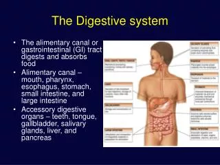
Digestive System Function: to obtain nutrients. Digestive System Function: obtaining nutrients. Activities of Digestion system: Ingestion – taking food or liquid into the mouth (eating or drinking). Movements Peristaltic and Segmentation Digestion Mechanical mastication

E N D
Digestive System Function: to obtain nutrients
Digestive System Function: obtaining nutrients • Activities of Digestion system: • Ingestion – taking food or liquid into the mouth (eating or drinking). • Movements • Peristaltic and Segmentation • Digestion • Mechanical • mastication • churning • Chemical • Enzymes and acids • Absorption – movement of substances into the body (across epithelium). • Elimination – involves compaction to eliminate waste from body
Layers of Gastrointestinal Tract • Four layers: • Know layers and their composition • Tunic mucosa • Epithelial • Propria • Muscularis mucosae • Tunic submucosa • Tunica muscularis externa • Tunica serosa-visceral peritoneum
Tunic Mucosa • Lines digestive tract • Moistened by glandular secretions • Simple or stratified depending on area of tract • Pleated for expansion (Surface Area)
1. Tunic Mucosa • A Mucous membrane • 1) Epithelium • 2) Lamina propria • 3) Muscularis mucosae 2. Tunic Submucosa • Areolar Connective Tissue • Innervation • May have glands
Muscularis Mucosa • Smooth muscle layer capable of plasticity • Ability to tolerate stretching • Visceral smooth muscle
3. Tunica Muscularis Externa • Smooth muscle layers • 1) Inner Circular Layer • 2) Outer Longitudinal Layer 4. Tunica Serosa (or Adventitia*) Serous membrane – visceral peritoneum • * Name depends on location: • Inside peritoneal cavity = serosa • Outside peritoneal cavity = adventitia
Serous Membranes • The Peritoneum: Two layers • Visceral peritoneum (a.k.a serosa) • Parietal peritoneum • Lines inner surfaces of body wall • Mesenteries: Fused double sheets of peritoneal membrane – to suspend portions of digestive tract: • Greater omentum • Lesser omentum • Mesentery proper • Transversemesocolon • Sigmoid mesocolon
Retroperitoneal Structures – these are attached to posterior abdominal wall • Ascending colon • Descending colon • Duodenum • Pancreas
Salivary glands • Parotid • Sublingual • Submandibular • Slightly different secretions • Release enzymes - amylase • Lubrication oral cavity
Fig 25.6
Fig 25.7 Only example of gomphosis joint Incisors -clipping/cutting Canines-tearing/slashing Premolars- mashing/grinding Molarsmashing/grinding
Teeth Regions: Crown Neck Root Layers: Enamel Dentin Pulp Cavity with Pulp Cementum Apical foramen Root canal Periodontal membrane
Voluntary control Pharynx and Esophagus Deglutition Swallowing Epiglottis closes over larynx
Tunica muscularis has three layers of muscle Fig 25.11 Tunica mucosa has folds, rugae when empty
Histology of Gastric glands Chief - Pepsinogen; Parietal - HCl; Neck/Mucous Cells • Secretin and cholecystokinin • Inhibit gastric secretion
Small Intestine Duodenum Jejunum Ileum Increase Surface Area for Absorption Plicae Circulares Intestinal villi Microvilli • Lacteal (terminal lymphatic) • for lipid absorption • Intestinal glands • Goblet cells • Stem cells
Diagrammatic view highlighting the distinguishing features of each region of the small intestine.
Duodenum: • Duodenal (Brunner’s) glands produce: • secretin • CCK • Alkaline mucus
Large intestine • Functions of large intestine • Reabsorb water and compact feces • Absorb vitamins • Store fecal matter • Cecum • Ileocecal valve • Collects material from small intestine • Vermiform appendix • Colon - ascending, transverse, descending and sigmoid • Rectum
Histology of Large Intestine Large Intestine: Lack of villi Abundance of goblet cells Mucous-secreting intestinal glands Muscularis reduced to Taenia coli Fatty appendices
Other digestive organs Horizontal section through the upper abdomen showing the position of the liver relative to other visceral organs.
Pancreas Exocrine: acini Endocrine: Pancreatic islets Isles of Langerhans