Download
1 / 52
540 likes | 549 Vues
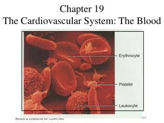
19 Blood. NOTE: Presentations extensively modified for use in MCB 244 & 246 at the University of Illinois by Drs. Kwast & Brown (2011-2012). Chapter 19 Learning Objectives. Describe the components and major functions of blood. Describe the composition and function of plasma.

E N D
19 Blood NOTE: Presentations extensively modified for use in MCB 244 & 246 at the University of Illinois by Drs. Kwast & Brown (2011-2012)
Chapter 19 Learning Objectives • Describe the components and major functions of blood. • Describe the composition and function of plasma. • List the characteristics and functions of erythrocytes, including hemoglobin, and describe erythrocyte formation (erythropoiesis) and recycling. • Explain the basis of blood typing and histocompatibility. • Describe the structure function of white blood cells and their formation. • Describe the structure/function of platelets. • Describe the mechanisms involved in blood clot formation.
Introduction to the Cardiovascular System • Cardiovascular System = blood, heart & blood vessels • Circulatory System = cardiovascular & lymphatic systems • Blood – a fluid connective tissue with matrix (plasma) and formed elements (cells) involved in: 1. Transport of dissolved substances (gases, nutrients, hormones, wastes) 2. Regulation of pH and ion composition 3. Restriction of fluid losses at injury sites (clotting) 4. Defense against toxins and pathogens (leukocytes) 5. Stabilization of body temperature
19-1 Physical Characteristics of Blood Fig. 19-1 pH = 7.4 Temp. = 38ºC Vol. = 4 - 6 liters (~7% body wt.) High Viscosity Fractionate Hemopoiesis: Hemocytoblasts → myeloid & lymphoid stem cells
19-2 Blood Fractions: Plasma • Makes up 50–60% of blood volume • More than 90% of plasma is water • Plasma and Interstitial Fluid are both extracellular fluids: exchange H2O, ions and small solutes (no protein) across the capillary wall • Composition of plasma and interstitial fluid differ in terms of • Levels of O2 and CO2 • Concentrations and types of dissolved proteins
19-2 Blood Fractions: Plasma Proteins • Albumins (60%) • Transport substances such as fatty acids, thyroid hormones, and steroid hormones; made by liver; major contributor to plasma osmotic pressure • Globulins (35%) • Antibodies, also called immunoglobulins • Transport globulins (small molecules): hormone-binding proteins, metalloproteins, apolipoproteins (lipoproteins), & steroid-binding proteins • Fibrinogen (4%) • Molecules that form clots and produce long, insoluble strands of fibrin • After soluble fibrinogen is turned into solid fibrin, remaining liquid portion = serum. • Other Plasma Proteins (1%) • Enzymes, hormones, prohormones: Composition fluctuates • Origin of Plasma Proteins: • 90% liver; Antibodies – plasma cells; Peptide hormones – endocrine organs
19-3 Blood Fractions: Red Blood Cells • Red blood cells (RBCs): 99.9% of formed elements in blood • Hemoglobin • The red pigment that gives whole blood its color • Binds and transports oxygen (and carbon dioxide) • Red blood cell count: the number of RBCs in 1 microliter of whole blood • Male: 4.5 – 6.3 million; Female: 4.2 – 5.5 million • Hematocrit (HCT or Ht) (packed cell volume [PCV] also used): percentage of RBCs in centrifuged whole blood • Male: 40 – 54; Female: 37 – 47
19-3 Blood Fractions: Red Blood Cells Figure 19–2 • Erythrocyte Structure – biconcave disc • High surface-to-volume ratio • Quickly absorbs and releases oxygen • Discs form stacks called rouleaux • Smooth the flow through narrow vessels • Discs bend and flex to enter small cpaillaries: • 7.8 µm RBC passes through 4 µm capillary Figure 19–2d
19-3 Blood Fractions: Red Blood Cells • Lack organelles including nuclei, mitochondria & ribosomes • No cell division or repair possible • Anaerobic metabolism only (no mitochondria) • Live ~ 120 days • Cell is 97% hemoglobin (Hb) • ~280 million Hb/RBC, 4 O2 binding heme/Hb = >1 billion O2/RBC x 25 trillion RBCs/indiv. = > 25 x 1021 O2/indiv.! • 1/3 of all cells (25 trillion out of 75 trillion) are RBCs! • Normal hemoglobin 12 – 18 g/dL whole blood
19-3 Blood Fractions: Red Blood Cells • Hemoglobin Structure/Function • Complex quaternary structure • 2 α chains & 2 β chains: • Each chain has 1 molecule of heme with O2 binding iron • Oxyhemoglobin(O2 bound, bright red) • Deoxyhemoglobin(no O2,burgundy) • 98.5% of O2 carried by Hb compared to 20% of CO2; the latter is bound to amino acids on α and β chains NOT heme – called carbaminohemoglobin • At peripheral capillaries, low plasma O2 leads to release of O2 and binding of CO2 • At lungs, opposite occurs - O2 loaded and CO2 expelled Figure 19–3
19-3 Blood Fractions: Red Blood Cells Hemoglobin Oxygen Dissociation Curves Comparison of Fetal vs. Adult Hb and Effects of pH, temp and di- (or bis-) phosphoglycerate (DPG or BPG) Fig. 23–22
19-3 Blood Fractions: Red Blood Cells • Hemoglobin Disorders: • Anemia = O2 starvation due • to: • Insufficient numbers of • erythrocytes • 2. Low hemoglobin • 3. Abnormal hemoglobin Fig. 19-4 • Thalassemia = inability to produce sufficient α or β chains → • slow RBC production and fragile & short-lived cells • Sickle-Cell Anemia = single amino acid mutation in β chain • When Hb highly oxygenated, cells have normal shape • When Hb O2 low, adjacent Hbs interact and RBCs deform into • crescent shape: cells become fragile and block capillaries
19-3 Blood Fractions: Red Blood Cells • RBCTurnover • 1% of circulating RBCs “wear out” every day (lack repair mechanisms) • That’s about 3 million RBCs/second! • Macrophages of liver, spleen and bone marrow monitor RBCs and engulf before membranes rupture (hemolyze) • Phagocytes break hemoglobin into constitutive components: • Globular proteins into amino acids; heme to biliverdin & iron • Transferrin, a plasma protein, transports iron back to bone marrow for new RBCs; excess transferrins removed by liver and spleen and iron stored as ferritin or hemosiderin • If excess hemoglobin breakdown, products can appear in urine; Hemoglobinuria • If whole red blood cells appear in urine, signals kidney or tissue damage; Hematuria
19-3 Blood Fractions: Red Blood Cells • Biliverdin (green) is converted to bilirubin (yellow) and is released into blood, filtered by liver, and excreted in bile • Jaundice= failure of bilirubin to be excreted in bile, collects in peripheral tissues → yellow skin & eyes • In gut, bilirubin → urobilins (yellow) & stercobilins (brown) via intestinal bacteria • urobilins absorbed or excreted in urine (hence yellow color to urine) • stercobilins remain in feces (hence brown color)
19-3 Blood Fractions: Red Blood Cells Fig 19–5 Recycling of Red Blood Cell Components
19-3 Blood Fractions: Red Blood Cells • Erythropoiesis (RBC Formation) • In adults, occurs only in myeloid tissue (red bone marrow): 1. Hemocytoblast differentiates into myeloid stem cell 2. Myeloid stem cell undergoes multiple stages of differentiation involving protein synthesis 3. Cell fills with Hb, loses organelles (nucleus too) 4. After ~ 4 days, reticulocytes are formed (Hb+ some ribosomes); remain in bone marrow for 2 days and then released into blood, where they account for 0.8% of total blood RBCs 5. After ~ 24h in circulation, they lose ribosomes (no more protein synthesis) = mature erythrocyte • Vitamin B12 necessary for stem cell division (also need B6 and folic acid) • B12 deficiency → pernicious anemia Fig 19–6
19-3 Blood Fractions: Red Blood Cells • Stimulating Hormones • Erythropoietin (EPO): hormone, released by kidney during hypoxia (low O2, e.g., high altitude or disease), anemia, ischemia, etc. • Stimulates RBC Production by: ↑ cell division rates (10x; ~30 million cells/s) ↑ Hb synthesis = ↓ maturation time • “Blood Doping” = injecting EPO or RBCs to enhance athletic performance: ↑ O2to tissues, but also ↑ hematocrit/viscosity → clots, stroke, heart strain, kidney failure
19-4 Blood Typing • All cell membranes have surface antigens: indicate “self” (antigen = substance that triggers immune response) • RBCs have 50+ surface antigens (e.g., glycoproteins or glycolipids) of which 3 are important for blood transfusion: agglutinogensA, B, Rh (D) • Blood types: A, B, AB or O • genetically determined by presence or absence of surface antigens (A, B & Rh) Rh+ = antigen Rh (85%); Rh- = none (15%) • At birth, blood contains antibodies against A and/or B antigens; however, this is not true for Rh as only sensitized individuals have antibodies to Rh (D) 40% 10% 46% 4%
19-4 Blood Typing • Transfusion Cross-Reactions • Plasma antibody meets specific surface antigen • Blood will agglutinate and hemolyze • Occur if donor and recipient blood not compatible O- = universal donor (AB universal recipient) • Cross-Match Testing: • tests for other antigens by reacting donors RBCs to recipients plasma Fig 19-7 Fig 19–8 Blood Type Test Anti-D+ = Rh+
19-4 Blood Typing: Rh antibodies Antibodies against Rh (D) antigen only form upon exposure and are small enough to cross placenta (unlike anti-A and anti-B antibodies) Hemolytic disease of newborn/Erythroblastosis fetalis: Rh- mom pregnant with Rh+ baby, gets exposed to D antigen during birth, makes anti-D antibodies. Pregnant with second Rh+ baby, her antibodies cross placenta, causing agglutination and lysis of fetal RBCs → anemia and death - fetal transfusion may be given and/or early delivery Easily prevented if known by treating mom with RhoGam (anti-Rh antibodies) during last 3 months of her first Rh+ pregnancy: prevents antibody formation in the first place. • Figure 19–9
19-5 Blood Fractions: White Blood Cells • Leukocytes (5 types) • Have nuclei & organelles but no hemoglobin (hence “white” or buff) • 5000 – 10,000 leukocytes/μl blood; < 1% total blood volume • Use blood to travel; most are found in connective tissue & lymph • Functions: • 1. Defend against pathogens • 2. Remove toxins and wastes • 3. Attack abnormal/damaged cells • Characteristics: • 1. Ameoboid movement – flow of cytoplasm into cellular processes • 2. Diapedesis (move out of blood): • a. Margination = adhere to vessel • b. Emigration = pass between endothelial cells • 3. Exhibit positive chemotaxis - pathogens, damaged tissue, other WBCs • 4. Phagocytosis (3 of 5) engulf pathogens and debris
19-5: 5 Types of Leukocytes a-d: Nonspecific defense e: Specific defense Fig 19–10 a-e White Blood Cells
19-5 White Blood Cells: A. Neutrophils or Polymorphonuclear [PMN] Leukocytes • Non-specific, defense • Phagocytic • 50-70% of all WBCs • 2-5 lobed nucleus • 12μm diameter • Granules (lysosomes) contain digestive enzymes & defensins that kill bacteria, fungi & enveloped viruses • Very mobile: first at injury • Life span < 10h • Functions: 1. Respiratory burst: H2O2& O2-, acts as bactericide 2. Degranulation: defensins (peptide) lyse bacteria 3. Prostaglandins: induce inflammation to stop spread of injury 4. Leukotrienes: attract phagocytes
19-5 White Blood Cells: B. Eosinophils or Acidophils • Non-specific defense • Phagocytic (2o) • 2–4% of circulating WBCs • Bilobed nucleus • 12 µm diameter; 9-day life • Functions: 1. Attack antibody-coated objects (bacteria, protozoa, cell debris) 2. Defense against large parasites • Excrete toxic compounds (1o) • Nitric oxide (NO) & Cytotoxic enzymes • Sensitive to allergens • Control inflammation with enzymes that counteract inflammatory effects of neutro- philsand mast cells eosin dye
19-5 White Blood Cells: C. Basophils • Non-specific defense • Not phagocytic • < 1% of WBCs • “U” shaped nucleus • 8-10μm diameter • Granules contain histamine– dilate blood vessels heparin – prevent clotting • Life span = 9 d • Similar in function to mast cells in tissue; enhance their function • Functions: 1. Inflammation 2. Allergic response (via histamine)
19-5 White Blood Cells: D. Monocytes • Non-specific defense • Phagocytic • 2-8% of WBCs • Kidney shaped nucleus • 15μm + diameter • Circulate 24 h, then exit to tissues = macrophage Life span = several months • Functions: 1. Phagocytosis: viruses and bacteria 2. Attract phagocytes 3. Attract fibroblasts for scar formation 4. Activate lymphocytes: to mount immune response
19-5 White Blood Cells: E. Lymphocytes • Immune-Specific Response • 20-30% of WBCs • Large round nucleus • 5-17μm diameter • Migratory between blood and tissues (bidirectional) • Most in lymphatic system • Life span = days to lifetime • Function (depends on type [3]): 1. T cells: cell-mediated immunity (attack foreign cells directly or control the activity of other lymphocytes) 2. B cells: humoral immunity (differentiate into plasma cells & synthesize and secrete antibodies) 3. Natural Killer (NK) cells: immune surveillance (detect and destroy abnormal tissue; e.g., cancer)
19-5 Leukocyte Disorders & Diagnostics • Changes in differential count and WBC profiles can signal infections, inflammation, and allergic reactions • Leukopenia: low WBC count • Leukocytosis: high WBC count normal infection ↑ WBCs from 7,500 - 11,000/μl >100,000/μl → leukemia, cancerous stem cells, WBCs produced are immature and abnormal • Infectious Mononucleosis: Epstein Bar virus infection causes production of excess agranulocytes (monocytes and lymphocytes) that are abnormal
19-5 White Blood Cell Production: Leukopoiesis & Lymphopoiesis • All blood cells originate from hemocytoblasts, which produce: 1. Myeloid Stem Cells • Differentiate into progenitor cells, which produce all WBCs except lymphocytes 2. Lymphoid Stem Cells • Lymphopoiesis: the production of lymphocytes • All WBCs, except monocytes, develop fully in bone marrow; • Monocytes develop into macrophages in peripheral tissues
19-5 Leukopoiesis • Myeloid stem cells → Basophils, Eosinophils, Neutrophils, Monocytes as directed by specific colony stimulating factors (CSFs) produced by Macrophages and T cells • Different CSFs (hormones) results in different cell types: • M-CSF stimulates monocyte production • G-CSF stimulates production of granulocytes (neutrophils, eosinophils, and basophils) • GM-CSF stimulates granulocyte and monocyte production • Multi-CSF accelerates production of granulocytes, monocytes, platelets, and RBCs
19-5 Lymphopoiesis • Hemocytoblasts differentiates into Lymphoid Stem Cells → Prolymphocytes → Lymphocytes • Some lymphocytes are derived from lymphoid stem cells that remain in bone marrow → B cells and NK cells • Many lymphoid stem cells migrate to peripheral lymphoid tissues (e.g., thymus, spleen & lymph nodes) and then differentiate into mature lymphocytes • Lymphoid stem cells in the thymus give rise to T cells (Discussed in much greater detail in Chapter 22)
19-5 Origins & Differentiation of Blood Formed Elements Fig 19–11
19-6 Blood Fractions: Platelets • Flattened cell fragments involved in human clotting systems • No nucleus (non-mammalian vertebrates have whole cells involved in clotting called thrombocytes) (thrombo- = clot) • 2-4 µm diameter, 1 µm thick • Constantly replaced, removed by spleen (phagocytized) • 9–12 days in circulation • 150,00 - 500,000 / µl of blood • Thrombocytopenia – abnormally low platelet count • Thrombocytosis – high platelet count (infection, inflammation, cancer) • 1/3 are reserved (in spleen and other organs) for emergencies
19-6 Blood Fractions: Platelets • Three Functions of Platelets: • Transport & release important clotting chemicals • Temporarily patch damaged vessel walls (plug) • Actively contract tissue after clot formation (contain actin & myosin) • Platelet Production (Thrombocytopoiesis) • Megakaryocytes in bone marrow breaks off membrane-enclosed cytoplasm (each megakaryocyte can produce ~4000 platelets) • Induced by • 1. Thrombopoietin (TPO) from kidney • 2. Interleukin-6 (IL-6) – stimulates platelet formation • 3. Multi-CSF (promotes growth of megakaryocytes)
19-7 Hemostasis • Hemostasis is the cessation of bleeding • Consists of three complex (and not necessarily sequential or independent) phases: 1. Vascular phase 2. Platelet phase 3. Coagulation phase
19-7 Hemostasis: 1. Vascular Phase • A cut triggers vascular spasm that lasts 30 minutes • Three steps of the vascular phase 1. Endothelial cells contract: • expose basal lamina to bloodstream 2. Endothelial cells release: • chemical factors: ADP, tissue factor, and prostacyclin • local hormones: endothelins, which stimulate smooth muscle contraction and cell division 3. Endothelial plasma membranes become “sticky”: • seal off blood flow
19-7 Hemostasis: 2. Platelet Phase ▪ Platelet adhesion (attachment) – begins within 15 s of injury • Adhere to sticky endothelial surfaces, basal lamina & exposed collagen fibers ▪ Platelet aggregation (stick together) ▪ Forms platelet plug (closes small breaks) ▪ Activated platelets release clotting cmpds feedback): • ADP→ platelet aggregation • Thromboxane A2& serotonin → vascular spasm • Clotting factors • Platelet-derived growth factor → blood vessel repair • Calcium ions → aggregation
19-7 Hemostasis: 2. Platelet Phase • Platelet aggregation must be controlled and the area restricted. Several factors limit the growth of the platelet plug: • Prostacyclin: released by endothelial cells, inhibits platelet aggregation • Inhibitory compounds released by WBCs entering area • Circulating plasma enzymes - break down ADP at plug • Negative (inhibitory) feedback: e.g., serotonin blocks ADP • Development of blood clot - isolates and restricts the area
19-7 Hemostasis: 3. Coagulation Phase • Begins 30 seconds or more after the injury • Blood clotting (coagulation) • Cascade reactions: • chain reactions of enzymes and proenzymes • form three pathways (extrinsic, intrinsic & common) • convert circulating fibrinogen into insoluble fibrin • Clotting Factors • Also called procoagulants (Ca2+ & 11 different proteins) • Many of the proteins are proenzymes • Required for normal clotting Figure 19–12a
19-7 Hemostasis: 3.Coagulation Pathways 1. Extrinsic pathway • Begins in the vessel wall (endothelial cells), outside of bloodstream • Damaged cells release Factor III or tissue factor (TF) • TF + Ca2+ + clotting factor VII = enzyme complex that activates Factor X 2. Intrinsic pathway • Begins with circulating proenzymes, within bloodstream • Activation of enzymes (usually Factor XII) by collagen • Platelets release factors (e.g., PF–3) • Series of reactions then activates Factor X 3. Common pathway • Where intrinsic and extrinsic pathways converge • Forms enzyme prothrombinase • Converts prothrombin to thrombin • Thrombin converts fibrinogen to fibrin
19-7 Hemostasis: Convergence of Coagulation Pathways Figure 19–12 The Coagulation Phase of Hemostasis NOTE: Both the extrinsic and intrinsic pathways produce thrombin, but the extrinsic is shorter and faster; thus, it results in the production of a small amount of thrombin first that is then later reinforced by the intrinsic pathway.
19-7 Hemostasis: Other Considerations • Positive Feedback: Production of thrombin by common pathway stimulates formation of tissue factor (TF) and PF-3 from platelets, thus forming a positive feedback loop with both the intrinsic and extrinsic pathways, respectively • Clotting: Area Restriction – affected by factors that either deactivate or remove factors/agents: • Anticoagulants (plasma proteins) • Antithrombin-III • Alpha-2-macroglobulin • Heparin (produced by basophils and mast cells) • Protein C (activated by thrombomodulin) • Prostacyclin – inhibits platelet aggregation
19-7 Hemostasis: Other Considerations • Dietary: Calcium Ions and Vitamin K affect almost all aspects of clotting • Calcium ions (Ca2+) needed for all 3 pathways (intrinsic, extrinsic and common) • Vitamin K required by liver for synthesis of 4 of the clotting factors, including prothrombin • Clot Retraction (occurs after clot formation) • Platelets contract, pull torn area together and reduce size of damaged area (takes 30–60 min) • Fibrinolysis = slow process of clot dissolving • Thrombin and tissue plasminogen activator (t-PA): • activate plasminogen • Plasminogen produces plasmin, which digests fibrin strands
19-7 Bleeding Disorders • Thrombosis = clotting in undamaged vessels→ prevents or slows flow (intrinsic pathway) • Embolus = free floating thrombosis, blocks small vessels → tissue damage, heart attack, stroke • Disseminated Intravascular Coagulation: widespread clotting followed by systemic bleeding, rare: complication of pregnancy, septicemia or mismatched transfusion • Hemophilia = inadequate production of clotting factors • Type A → Factor VIII (X linked) • Type B → Factor IX • Type C → Factor XI