Download
1 / 32
390 likes | 649 Vues
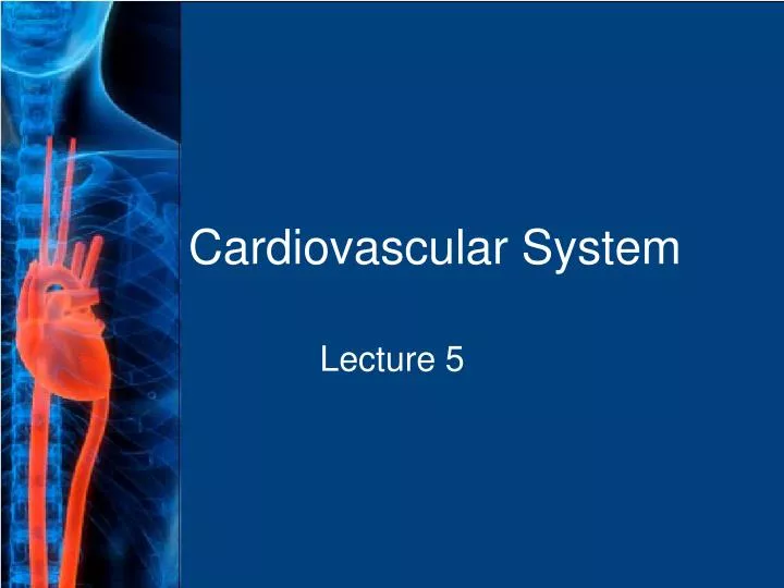
Cardiovascular System. Lecture 5. General. The cardiovascular system is a series of tubes and a muscular pump that provides a ONE-WAY street for blood, oxygen, and nutrients. Blood and nutrients travel through blood vessels (arteries, veins, and capillaries).

E N D
Cardiovascular System Lecture 5
General • The cardiovascular system is a series of tubes and a muscular pump that provides a ONE-WAY street for blood, oxygen, and nutrients. • Blood and nutrients travel through blood vessels (arteries, veins, and capillaries).
The cardiovascular system is fueled by a muscular pump called the heart. The heart is actually two pumps connected by a SEPTUM. • The right side of the heart pumps blood that is deficient in oxygen to the lungs [Pulmonary Circulation]. • The left side of the heart pumps blood that is rich in oxygen to the body [Systemic Circulation].
The Heart • The heart has three distinct layers of tissue. 1. endocardium - deepest layer. 2. myocardium - muscle 3. epicardium - outermost layer
The heart is a muscular organ that pumps blood and is enclosed in a membranous sac. This sac allows the heart to beat without friction. • This sac is called the PERICARDIUM. Peri- means “around”, cardium refers to the heart.
Peri/cardi/ectomy - sx procedure excising the pericardium. Peri/cardi/o/rraphy - suturing a wound of the pericardium. My/o/cardi/um - the muscular layer of the heart.
There are two sides of the heart. • There are two vertical divisions of the heart. • The top compartments are ATRIA • The bottom compartments are VENTRICLES • Therefore, there are right and left atria and right and left ventricles.
Left Atrium Right Atrium Left Ventricle Right Ventricle Apex Heart Chambers
Abbreviations for chambers: Right atrium RA Right ventricle RV Left atrium LA Left ventricle LV
The ventricles are larger than the atria. This is because the ventricles are responsible for pumping blood a farther distance than the atria. • Of the two ventricles, the left is larger than the right. This is because the left ventricle must pump blood to the entire body.
A rapid contraction of the atrium or ventricle is known as a FLUTTER. • Atrial flutter can cause chest pain and shortness of breath (SOB). • The rule for forming plural words from the singular that end in –um is to drop the –um and add an –a.
The prefix “tachy-” refers to rapid. a rapid heartbeat (pulse): tachycardia • The prefix “brady-” refers to slow. a slow heartbeat (pulse): bradycardia
Arteries bring blood AWAY from the heart. • Veins bring blood TOWARD the heart. • Arteries usually carry blood with much oxygen. • Veins usually carry blood with little oxygen. • The RIGHT ATRIUM receives blood from all tissues of the body through veins. This blood is oxygen poor.
The blood brought back to the heart comes from three sources: • SUPERIOR VENA CAVA (SVC) brings blood from the top part of the body. • INFERIOR VENA CAVA (IVC) brings blood from the lower part of the body. • CORONARY SINUS brings blood from the heart muscle. All three sources empty into the RIGHT ATRIUM.
Once inside the right atrium, the blood must travel to the right ventricle. In order to do this, it must pass through the TRICUSPID VALVE. • The function of all heart valves is to allow one way travel of blood. It would be dangerous to have blood backflow because of different oxygen concentrations.
Aorta Pulmonary Artery Pulmonary Veins Left Mitral Vena Cava Tricuspid Right Apex Heart Valves
Once inside the right ventricle, the blood passes through the PULMONARY SEMILUNAR VALVE into the PULMONARY ARTERIES. • The pulmonary arteries carry oxygen-deficient blood to the lungs. • Once inside the lungs, the blood vessels branch until they reach one cell layer thick. These CAPILLARIES combine with the ALVEOLI of the lungs for the exchange of oxygen and carbon dioxide.
The blood now has much oxygen. It returns to the heart by the PULMONARY VEINS. There are four pulmonary veins that empty into the LEFT ATRIUM. • The blood then must pass through the MITRAL VALVE (BICUSPID VALVE) into the left ventricle. • From the left ventricle the blood passes through the AORTIC SEMILUNAR VALVE in the AORTA. • The aorta is the largest artery of the body.
The contraction of the left ventricle sends blood rich in oxygen all over the body. There are three arteries that bring blood to the head, neck, and upper extremities. There is one major vessel that brings blood to the abdomen and lower extremities. • Arteries are the large vessels that bring blood away from the heart. These vessels branch into smaller ARTERIOLES which eventually branch into CAPILLARIES which are only one cell thick.
The primary responsibility for initiating the heartbeat is with the SINOATRIAL NODE. This is located on the posterior wall of the right atrium. • Once this electric current is generated, atrial muscle contracts forcing blood into the ventricles. Once this occurs the heartbeat moves to another region called the ATRIOVENTRICULAR NODE.
Once this occurs, the AV node sends electrical impulses through a series of BUNDLE BRANCHES ending in PURKINJE FIBERS that stimulate the ventricles to contract.
Blood Supply to Heart • Coronary Artery System • right coronary • left coronary • left anterior descending • circumflex
The Cardiac Cycle and Heart Sounds • The CARDIAC CYCLE is the events that occur in one complete heartbeat. • The cardiac cycle has 2 phases: 1. contraction of the heart: SYSTOLE 2. relaxation of the heart: DIASTOLE
The atria and ventricles have different functions during the cardiac cycle. • When the atrium are contracting, blood flows into the ventricles. Therefore, the ventricles have to be relaxing. • When the atria are in systole, the ventricles are in diastole. • Electrical activity of the heart can be measured by an electrocardiogram (EKG or ECG).
EKG’s are electrical tracings of each part of the cardiac cycle. • Each time a different part of the heart contracts, an electrical impulse can be recorded from different areas on the thorax.
QRS complex – signals activity of the Purkinje fibers and Bundle of His P wave – signals atrial contraction T wave - signals ventricular relaxation
Microcardia - small heart Cardiomegaly (megalocardia) - enlargement of heart Myocardial Infarction (MI) - heart attack Hypertension - high blood pressure
ATHEROSCLEROSIS is a form of ARTERIOSCLEROSIS and is characterized by an abnormal accumulation of fat and fibrous tissue (scarring) in a blood vessel. • This leads to a narrowing of the LUMEN which causes a decrease in blood flow to a part of the body. • This condition can lead to NECROSIS, or cellular death.
To prevent blood clots, patients may take an ANTICOAGULANT. These are agents that delay blood coagulation (clotting). • Anticoagulants are used to prevent THROMBUS formation (THROMBOGENESIS). • THROMBOLYSIS is accomplished with THROMBOLYTIC AGENTS or medications that destroy a clot.
An ANEURYSM is a weakened blood vessel wall caused by DILATION of the vessel. This causes the vessel to balloon and eventually burst. • There are two types of aneursym: 1. Fusiform – the wall dilates equally resulting in a tubular swelling. 2. Sacculated – a balloon is attached to the vessel by a narrow stalk.