Download
1 / 34
420 likes | 796 Vues
Mammalian Nervous System Chapter 46. How Is the Mammalian Nervous System Organized?. Vertebrate nervous systems consist of a brain, a spinal cord, and peripheral nerves that extend throughout the body. The central nervous system or CNS contains the brain and spinal cord.

E N D
Mammalian Nervous System Chapter 46
How Is the Mammalian Nervous System Organized? Vertebrate nervous systems consist of a brain, a spinal cord, and peripheral nerves that extend throughout the body. The central nervous system or CNS contains the brain and spinal cord. The peripheral nervous system or PNS consists of the cranial and spinal nerves that connect the CNS to all tissues.
Figure 46.1 Organization of the Nervous System Add Figure 46.1
Figure 46.2 Development of the Human Nervous System (Part 1)
Figure 46.2 Development of the Human Nervous System (Part 2) Add Figure 46.2 middle panel only (40 days)
Figure 46.2 Development of the Human Nervous System (Part 3) Add Figure 46.2 bottom panel only (100 days)
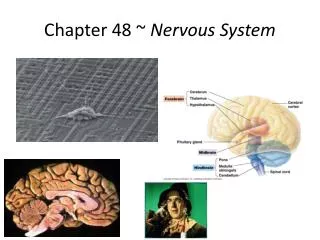
The three parts of the embryonic brain develop into structures in the adult brain. The hindbrain becomes the medulla, the pons, and the cerebellum. Physiological functions, such as breathing and swallowing are controlled by the medulla and pons. Muscle control is coordinated in the cerebellum.
The embryonic midbrain becomes structures that process visual and auditory information. Together the hindbrain and midbrain are known as the brain stem.
The embryonic forebrain develops the central diencephalon and the surrounding telencephalon. The diencephalon consists of the: • Thalamus, which is the final relay station for sensory information • Hypothalamus, which regulates physiological functions such as hunger and thirst
The telencephalon consists of two cerebral hemispheres and is also called the cerebrum. An evolutionary trend in which the telencephalon increases in size and complexity in vertebrates is telencephalization. In humans, the telencephalon is the largest brain region and is involved in sensory perception, learning, memory, and behavior.
The spinal cord: • Conducts information between brain and organs. • Integrates information coming from PNS. • Responds by issuing motor commands.
Anatomy of the spinal cord: • Gray matter is in the center, and contains cell bodies of spinal neurons. • White matter surrounds gray matter and contains axons that conduct information up and down the spinal cord. • Spinal nerves extend from the spinal cord.
Spinal reflex: afferent information converts to efferent activity without the brain. The knee-jerk reflex is monosynaptic: • Stretch receptors send axon potentials through dorsal horn to ventral horn, via sensory axons. • At synapses with motor neurons in the ventral horn, action potentials are sent to leg muscles, causing contraction.
Figure 46.3 The Spinal Cord Coordinates the Knee-Jerk Reflex
Structures in primitive regions of the telencephalon form the limbic system. • Amygdala: involved in fear and fear memory • Hippocampus: transfers short-term memory to long-term memory
Cerebral hemispheres are dominant in mammals. Cerebral cortex– a sheet of gray matter covering each hemisphere that is convoluted to fit into the skull • Gyri: (sing. gyrus) ridges of the cortex • Sulci: (sing. sulcus) valleys of the cortex
Regions of the cerebral cortex have specific functions. Association cortex is made up of areas that integrate or associate sensory information or memories. Four cortical lobes: • Temporal • Frontal • Parietal • Occipital
Temporal lobe: • Receives and processes auditory information • Association areas of the temporal lobe involve: • Identification • Object naming • Recognition Agnosia: a disorder of the temporal lobe
Frontal Lobe: • Central sulcus: divides the frontal and parietal lobes • Primary motor cortex is located in front of the central sulcus and controls muscles in specific body areas. • Association areas involve: • Planning • Personality
Parietal Lobe: • Primary somatosensory motor cortex is located behind the central sulcus; it receives touch and pressure information. • Association areas involve attending to complex stimuli. Contralateral neglect syndrome: an inability to recognize stimuli on one side of the body when the opposite parietal lobe is damaged
Occipital Lobe: • Receives and processes visual information • Association areas involve: • Making sense of the visual world • Translating visual experience into language
Autonomic Nervous System (ANS): the output of the CNS that controls involuntary functions ANS has two divisions that work in opposition: one will increase a function and the other will decrease it. Sympathetic and parasympathetic divisions are distinguished by anatomy, neurotransmitters, and their actions.
Sympathetic and parasympathetic divisions have different anatomy. The sacral region contains preganglionic neurons of the parasympathetic region. The thoracic and lumbar regions contain sympathetic preganglionic neurons.
Electroencephalogram (EEG): • Measures activity of groups of neurons • Records changes in electrical potential between electrodes, over time Electromyogram (EMG) records skeletal muscle activity. Electrooculogram (EOG) measures eye movement.
Figure 46.14 Patterns of Electrical Activity in the Cerebral Cortex Characterize Stages of Sleep (1) Add Figure 46.14 (A)
Figure 46.14 Patterns of Electrical Activity in the Cerebral Cortex Characterize Stages of Sleep (2)
Language areas: • Broca’s area located in the frontal lobe: damage results in slow or lost speech but a person can read and understand language. • Wernicke’s area is in the temporal lobe: damage results in an inability to speak sensibly, as written or spoken language is not understood. A person may still be able to produce speech. • Angular gyrus: adjacent area essential for integrating spoken and written language
Figure 46.16 Imaging Techniques Reveal Active Parts of the Brain