Download
1 / 9
90 likes | 99 Vues
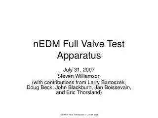
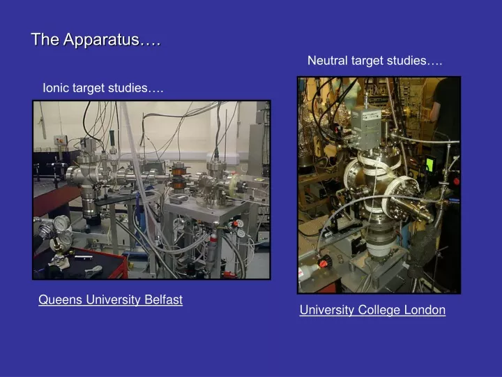
The Apparatus…. Neutral target studies…. Ionic target studies…. Queens University Belfast. University College London. Schematic of the Ion Beam Apparatus. Primary Beam Collector. Charged Fragment Detector. Extraction and Focussing Lenses - ions are accelerated to 1-2 keV. Einzel lens.

E N D
The Apparatus…. Neutral target studies…. Ionic target studies…. Queens University Belfast University College London
Schematic of the Ion Beam Apparatus Primary Beam Collector Charged Fragment Detector Extraction and Focussing Lenses -ions are accelerated to 1-2 keV Einzel lens Deflection Plates 45o Parallel Plate deflectors Laser Beam Ion Source -ions produced via discharge Interaction Region Neutral Fragment Detector Selection Magnet
Plasma Discharge Ion Source - creates a plasma of ions and electrons In the fringe region between the anode and cathode, electrons are stripped off the introduced gas by the strong electric field. The electrons then spiral due to the surrounding magnetic field thus colliding with more atoms ionizing them. The ions produced are self-extracted through the aperture due to the electric field distribution and are then focused and transported.
Mass Selection Magnet - used to select the particular ion species to study. Ions passing through the magnet field region are deflected due to the Lorentz force. For a particular magnetic field setting, ions of different mass/charge ratios are deflected through different angles. By varying the magnet field we can select a particular mass/charge ratio ion to exit through the final aperture.
Interaction Region - interaction of ions with focused laser beam. The laser system used is the RAL Astra 790nm Ti:Sapphire laser giving 50fs, 25mJ pulses at 10Hz. When focused with a relatively soft focusing lens, we obtain intensities between 1016 – 1017 Wcm-2. The ionized ions are carried along with the initial momentum of the primary ion beam. A set of plates can also be used to bias the interaction region if needed.
Parallel Plate Analyser - used to separate product ions from primary ion beam. An electric field is applied across the 45o parallel-plate region. Ions of higher charge/mass ratio are deflected more by the electric field thus one can separate out the ionized products from the primary ion beam. The main ion beam is dumped in a well-baffled Faraday cup whilst the product ions are detected by a CEM. An on-axis CEM is used to detect any neutral fragments from molecular studies.
Focussed Laser Beam Neutral Target Apparatus The gas is effused into the interaction region via a hypodermic needle. The focused laser interacts with the gas target and the product ions are extracted by a weak electric field across the interaction region. After drifting up the drift-tube region, the product ions are detected by a pair of microchannel plates with their time-of-flight recorded. The system can be operated in Spatially-resolved geometry mode or Wiley-McLaren mode.
focussed laser direction y x z ion beam direction Scanning Techniques…. Intensity Scan With the Intensity scan, interact the ion beam with the centre of the laser focus. Then vary the peak focus intensity by varying the input power of the laser. Thus measure Ion yield (ionization) as a function of laser intensity.
focussed laser direction y x z ion beam direction Scanning Techniques…. Intensity Selective Scan With the Intensity Selective Scan, drive the laser focus through the ion beam. By interacting with different intensity slices of the focus, we again measure the Ion yield as a function of intensity. This technique has the added advantage in that the low intensity regions are enhanced by increased volume.