Download
1 / 29
290 likes | 412 Vues
Immune System. The influenza epidemic of 1918-1919 killed 22 mil-lion people in 18 months. With 25 million American infected, the Red Cross often worked around the clock. 3 million people will die from malaria this year. .

E N D
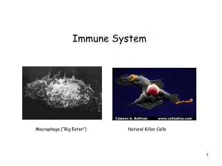
Immune System
The influenza epidemic of 1918-1919 killed 22 mil-lion people in 18 months. With 25 million American infected, the Red Cross often worked around the clock. 3 million people will die from malaria this year.
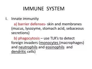
Innate immunity is one that you are born with and does not require the exposure to the disease to activate it. First line of defense-This defense works to keep the pathogen out of the body. Integuments System- -skin & mucous membranes -contains keratin which is an enzyme resistant to many bacterial enzymes -skin's fatty acids and secretion from sweat and oil glands are toxic to bacteria
Body passages-the mucous produced by the respiratory system traps microorganisms and the cilia sweeps it out The first line of defense in the respiratory tract (trachea). The orange cells produce mucus that that traps microorganisms that enter. The yellow cells are ciliated which beat in unison to expel mucus and trapped microorganisms upward to pharynx.
Stomach- the pH kills ingested bacteria. Tears contain lysozymes which destroy cell walls of bacteria Urine flushes bacteria out. Natural fauna of our own bacteria are harmless can out compete other bacteria.
This is skin that has been cleaned with soap and rinsed clean. Even with this treatment, skin cells may be home to million of harmless bacteria (green rod-shaped cells). Second line of defense of innate immunity I. Non-specific- direct; immediate invader can be anything: • Chemical Response- The injured cells release chemicals to help destroy invaders • 1. Histamine- Mast cells are cells found lining nasal passages and in connective tissue, and basophile cells are leukocytes are circulating in the body. When antigens such as pollen attach to these cells, they are damaged and release histamine. Histamine is a chemical that causes a.) vasodilatation b.) increase permeability of capillary beds. This causes the area to become red and swollen.
The picture on the left is ragweed pollen. The other picture are mast cells being attacked by antigens. They will lyse and release histamine. Antihistamines will suppress this response. 2. Kinins (or chemokines)- polypeptides a.) increase circulation and capillary permeability b.) attract leukocytes to site of injury c.) affect nerve cells making area tender
3. Complement proteins (approx. 20 proteins) a.) Directly- form protein rings on bacteria membranes forming pores, water rushes in, bursts cells
b.) Indirectly- The complement protein coats the surface bacteria. There are recognition sites on the phagocytes that bind with proteins coating the bacteria. The phagocytes engulf the bacteria in a process called opsonization- 4. Interferon- is made and attaches to surface receptors on other cells making harder for viruses to attack.
B. Innate immunity-Second line of defense. Nonspecific Cellular-Uses leukocytes (white blood cells made in bone marrow). These include phagocytes, basophiles and natural killer cell which is a lymphocytes. 1. Phagocytes are commonly found in connective tissue that help form the lining of the many organs (i.e. liver, lungs and kidneys), and they are found circulating in the blood and lymph nodes. Phagocytes go to the site of infection and start engulfing bacterial cells and cellular debris. Pathogens are surrounded by a membrane. The vacuoles fuse with lysosomes to destroy the pathogens.
The lysosomes contain hydrolytic enzymes that destroy the bacteria. Phagocytes can literally eat themselves to death. Pus are the remains of phagocytes that have been destroyed.
*Types of Phagocytes -Eosinophils- weakly phagocytic cells attracted to area, kills invaders that have been clumped together. Will also kill parasitic worms by depositing toxic granules in them. -Neutrophils- first to arrive; numerous (1 billion made each day); survive only a few days. Expendable.
-Monocytes arrive- transform into large macrophages; very efficient also involved in primary immune response. A monocyte that has been transformed into a macrophage. This macrophage is engulfing bacterial cells and destroying them.
-Basophiles are leukocytes containing histamines. These are release when these circulating cells are damaged.
A natural killer cell (in the fore-ground) is killing a cancer cell by punching holes in the plasma membrane. Water will rush in and lyse the cell. -Natural killer cells are (lymphocytes) roam the body contacting cells looking for infected cells by viruses and/or cancer cells. Eukaryotic cells are transformed when infected by viruses and cancer cells are also transformed. Natural killer cells can differentiate these cells and will destroy these cells. It destroys cells by lysing the cells.
A number of natural killer cells are attacking a cancer cell. Water will rush in and lyse the cell. In general phagocytes in mammals have the ability to distinguish pathogens because they contain receptor sites called Toll-like receptors (TLR). TLR recognizes certain molecules that are common to pathogens but not mammals. -TLR4 found on phagocytes recognizes a lipopolysaccharide found on many bacteria. -TLR3 found inner membrane of vesicles from endocytosis recognizes DS RNA from viruses. TLR causes phagocytosis to occur. The vesicles then fuse with a lysosome. The lysosome releases nitric oxide to kill pathogen and then releases hydrolytic enzymes to break pathogens down.
This illustrates the interaction of the cellular and chemical interaction of the nonspecific immune response to produce the inflammatory response. 1. Chemical such as hista-mine are released by mast cells and macrophages. 2. Histamine increases capillary permeability. The area becomes swollen. 3. Phagocytes leave the capillary bed attracted to the site . 4. They clean up pathogens.
Specific response-or immune response is slower and requires time. The lymphocytes arise in bone marrow. Then the lymphocytes either stay in the bone marrow or migrate to the thymus gland to mature and differentiate. The bone marrow and thymus are primary lymphoid tissue. Once mature and differentiated, the lymphocytes migrate to secondary lymphoid tissue where they remain and are later activated. Secondary lymphoid tissue include, adenoids, tonsils, lymph nodes, the spleen and Peyer's patches found on the small intestines.
Lymph vessels include lymph capillary beds connected to lymph veins. (No lymph arteries). These veins move lymph like circulatory veins. As the lymph is moved through veins, it is filtered by lymph nodes where pathogens are removed. Lymph is a clear fluid that
is clear fluid that is left over from the circulatory capillaries. Lymph veins work like circulatory veins. The fluid is filtered by the lymph nodes. The lymph is returned to the circulatory system dumping into vena cava near the heart. There are 2 types of lymphocytes involved in the immune response. When immature, the 2 cells are indistinguishable. T-cells mature in thymus and have a large amount of ribosomes. B-cells mature in red bone marrow and have a large amount of E.R. The first cell is an immature lymphocyte. The second cell is a mature B- cell with extensive E.R. The last cell is a mature T-cell with its extensive amount of ribosomes.
Mature B-cells secrete AB and some are memory cells used to prevent a second infection. They are involved in the humoral response. Mature T-cells are involved in the cell mediated response and also the humoral response.
Antibodies bind to epitopes on the surface of an antigen. In the above example, 3 different AB molecules react with 3 epitopes on the same large AG molecule Antibodies (AB) are special proteins secreted in response to the presence of foreign substance or antigen. Antigen (AG) can be nucleic acid, carbohydrates, protein. AG usually large, over 5000 Daltons. AG can be free floating OR attached to invading virus or bacterial cells. A particular molecule, virus or bacteria may activate many different AB to be made. The part of the AG that causes an AB to be synthesized is termed an antigenic determinant or epitope.
Antibodies are proteins made of 4 polypeptides. 2 identical, heavy, polypeptide chains and 2 identical, light polypeptide chains. A light chain is bonded to a heavy chain by disulfide bonds. The part of the AB that binds to the AG is found on the variable regions of the light and heavy chains. AB are made by B-cell lymphocytes.
At the end of the of the AB are 2 binding sites for AG. These ends are called the variable regions and are specific for particular AG. The other parts are constant regions and are the same for each type of AB.
Five Different Classes of AB or immunoglobulins Ig -IgG- monomer- found late in response crosses placenta -IgA- dimer and monomer form -IgM- pentamer- monomer form helps with virgin B cells -IgD- monomers found surface B cells -IgE- monomer found in area with connective tissue helps with release histamine
Not only do B cells make AB but they have AB on the surface of the plasma membrane that is used as receptor site. T-cells also have antibody like receptor sites. This T-cell receptor site has both a variable and constant region.
How Do AB Work 1. Agglutination-AB bind to several AG clumping them together so phagocytes can devour them 2. Neutralization-AB will coat the epitopes so that the pathogen cannot interact with the receptor sites of cells 3. Certain interaction with AB and AG trigger complement system, the complement proteins cause holes in invading cells, H2O goes in, cell bursts 4. Precipitation-AB will attach to soluble AG so that it will precipitate out and can be easily engulfed (phagocytosis).
4. Opsonization can occur- bacterial cells will have so many AG determinants that the AB may coat the bacteria cell- the constant regions stick out- called the FC region. The phagocytes have FC receptor, so the phagocyte literally rolls over the invader and engulfs it.