Download
1 / 27
280 likes | 461 Vues
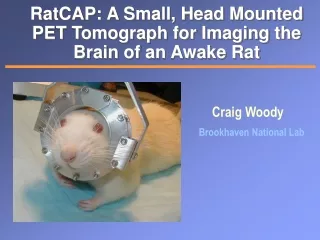
The RatCAP Conscious Small Animal PET Tomograph. Craig Woody Brookhaven National Lab. EuroMedIm 2006 Marseille, France May 10, 2006. Imaging The Awake Animal.

E N D
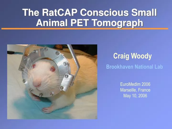
The RatCAP Conscious Small Animal PET Tomograph Craig Woody Brookhaven National Lab EuroMedIm 2006 Marseille, France May 10, 2006
Imaging The Awake Animal One wants to study neurophysiological activity and behavior in laboratory animals using PET in order to better understand these effects in humans. • Animals need to be anesthetized during PET imaging due to their inability to lie motionless in the scanner • Anesthesia can greatly depress brain functions and affect the neurochemistry that one is trying to study • Cannot study animal behavior while under anesthesia C.Woody,EuroMedIm 2006, 5/10/06
RatCAP: RatConscious Animal PET A miniature, complete full-ring tomograph mounted to the head of an awake rat. • Compact, light weight (< 200 g), low power detector • Small field of view (38 mm dia. x 18 mm axial) • Attached to the head of the rat and supported by a tether which allows reasonable freedom of movement for the animal C.Woody,EuroMedIm 2006, 5/10/06
Tomograph Ring Ring containing 12 block detectors of 2x2 mm2 x 5 mm deep LSO crystals with APDs and integrated readout electronics APD (Hamamastu S8550) LSO array Actual RatCAP Ring LSO Socket Readout chip APD C.Woody,EuroMedIm 2006, 5/10/06
Readout Electronics Custom ASIC (0.18 mm CMOS) 32 channels preamp, shaper, discriminator ~ 1W total power ZCD Bare chip Totally Digital Output 5 bit address Leading edge gives timing No ADC’s Minimizes cabling Packaged chip C.Woody,EuroMedIm 2006, 5/10/06
Energy Resolution Threshold scan Threshold (mV) Differential pulse height spectrum Threshold (mV) Energy resolution is used primarily for scatter rejection • Presently have common threshold for all • channels • Measure count rate as a function of • threshold for all channels and • differentiate spectra • Requires setting threshold low enough • for good efficiency for lowest channel • (~ 146 keV) poor timing resolution • for others • Next version of chip will have • independent gain and threshold • settings for each channel FWHM ~ 23% Thresh C.Woody,EuroMedIm 2006, 5/10/06
Timing Resolution 2t ~28 ns Timing resolution is used to reject randoms background Coincidence window 2t ~ 2 • FWHM • Large background from activity in the rat’s • body • High singles rate (~ 50 KHz/block) • Not possible to shield background due to • added weight • Next version of chip will use a leading edge • discriminator and time-over-threshold • measurement ( energy) for time walk • correction and improved timing resolution C.Woody,EuroMedIm 2006, 5/10/06
Position Resolution RatCAP 7 mm 15 mm FWHM (mm) R4 MicroPET Point Source Resolution 3.4:1 activity ratio (striatum to background) Rat Brain Striatum Phantom Intrinsic Spatial Resolution 1.28 mm FWHM Concorde P4 MicroPET = 1.75 mm UCLA MicroPET = 1.58 mm C.Woody,EuroMedIm 2006, 5/10/06
Sensitivity Count Rate (kcps) 4 mCi 2t = 40 ns Small Animal PET Sensitivities (Threshold = 250 keV) microPET (original) 0.56% ATLAS 1.8% microPET R4 4.4% microPET P4 2.3% microPET II (proto) 2.3% microPET Focus 220 3.4% microPET Focus 120 7.7% microPET R4 = 45 kcps @ 6 uCi/cc RatCAP Point Source Sensitivities 0.7% @ 150 kev 0.4% @ 400 keV C.Woody,EuroMedIm 2006, 5/10/06
RatCAP Support System Weight is completely counterbalanced (animal feels only inertia) Inner ring attaches to head which mounts to tomograph Gimbal ring allows head movement C.Woody,EuroMedIm 2006, 5/10/06
Adaptation to Wearing the Ring Corticosteroid Levels in Untrained rats wearing the RatCAP Animal training and conditioning should reduce stress levels significantly C.Woody,EuroMedIm 2006, 5/10/06
Animal Training C.Woody,EuroMedIm 2006, 5/10/06
Mounting the RatCAP to the Head C.Woody,EuroMedIm 2006, 5/10/06
Rat wearing the RatCAP C.Woody,EuroMedIm 2006, 5/10/06
First Images 517 g rat, 802 uCi 18F-FDG i.p. injection 45 min awake uptake, then chloral hydrate euthanasia MicroPET R4 scan • 10 min, LLD = 250 keV, 2 = 10 ns • 3D MLEM (20 iterations) RatCAP scan • 33 minutes livetime over 150 min scan • equivalent to 1.9 X decays of R4 scan • Monte Carlo-based 3D MLEM • 200 iterations • Randoms correction • No efficiency correction (yet) MicroPET R4 RatCAP Overlay RatCAP FOV C.Woody,EuroMedIm 2006, 5/10/06
Methamphetamine Images Using the RatCAP RatCAP vs MicroPET Time Activity Curve 250 200 150 ROI Activity in nCi/cc RatCAP MicroPET 100 50 0 0 100 200 300 400 500 600 700 Time in seconds RatCAP MicroPET The resolution of the RatCAP is slightly better than the commercial MicroPET scanner C.Woody,EuroMedIm 2006, 5/10/06
Fluoride Scan Brain Artifact ( due to randoms correction) Skull 3 mCi 18F Injection Uptake mainly in the bone C.Woody,EuroMedIm 2006, 5/10/06
Summary • The ability to image the awake animal will open up many new possibilities in neurophysiology and neurochemistry • The RatCAP is a fully functional miniature 3D tomograph that can be used for PET imaging of live, unanesthesized rats, and will provide one of the first opportunities to perform detailed studies on awake animals • The device can also be used as a standard small animal tomograph for anesthesized animals, and can be used for other applications using the same detector components • The first preliminary studies using the RatCAP have been completed, and we are now looking forward to the first real awake animal images and to improving its design in the future. C.Woody,EuroMedIm 2006, 5/10/06
The Team P. Vaska, C. Woody, D. Schlyer, J.-F. Pratte, P. O’Connor, V. Radeka, S. Shokouhi, S. Stoll, S. Junnarkar, M. Purschke, S.-J. Park, S. Southekal, V. Dzhordzhadze, W. Schiffer, D. Marsteller, D. Lee, S.Dewey, A. Villanueva, S. Boose, A. Kandasamy, B. Yu, A. Kriplani, S. Krishnamoorthy, S. Maramraju Brookhaven National Laboratory J. Neill, M. Murphy, T. Aubele, R. Kristiansen Long Island University R. Lecomte and R. Fontaine University of Sherbrooke C.Woody,EuroMedIm 2006, 5/10/06
Backup slides C.Woody,EuroMedIm 2006, 5/10/06
Effects of Anesthesia • Blood flow and metabolism depend on type and dose of anesthesia • Global and regional cerebral blood flow is affected by anesthesia • Cerebrovascular reactivity to CO2 is perturbed • Neural activity is suppressed • Effects are different for different animals and species M.Pomper, Johns Hopkins From the 9th International Conference: Peace Through Mind/Brain Science, Hamamatsu, Japan, Jan. 30-31, 2002 The effect of anesthesia on the uptake of β-CFT on dopamine transporters in the monkey brain. C.Woody,EuroMedIm 2006, 5/10/06
Effects of Anesthesia • Blood flow and metabolism depend on type and dose of anesthesia • Global and regional cerebral blood flow is affected by anesthesia • Cerebrovascular reactivity to CO2 is perturbed • Neural activity is suppressed • Effects are different for different animals and species Reduction in glucose metabolism with isoflurane in humans Similar effects are seen in the rat M.Pomper, Johns Hopkins D.B. Stout et al, UCLA 1998 C.Woody,EuroMedIm 2006, 5/10/06
Neurotransmitter Activity in the Brain MAO A DA DA 11C-Cocaine DA DA DA DA DA DA DA DA DA DA DA DA DA DA DA DA DA DA signal Drugs like cocaine can block the re-uptake sites for neurotransmitters like dopamine which upsets the normal equilibrium and can cause effects of addiction C.Woody,EuroMedIm 2006, 5/10/06
Time Signal Processing Module FPGA time stamps and packages singles events into 64 bit words 1.3 ns bit resolution VME data acquisition VxWorks running PDAQ Up to 40 MB/s to Linux box Online monitoring Singles & coincidence rates Offline processing Time and energy calibrations Coincidence sorting Randoms estimation Efficiency normalization Sinogram sorting Readout System FPGA C.Woody,EuroMedIm 2006, 5/10/06
Rat wearing the RatCAP C.Woody,EuroMedIm 2006, 5/10/06
Wrist Scanner for Measuring the Arterial Input Function Obective: to perform non-invasive quantification of the Arterial Input Function using PET Imaging. Activity in the surrounding veins produce a significant background which can be rejected using the good spatial resolution from PET C.Woody,EuroMedIm 2006, 5/10/06
Wrist Phantom Studies Artery Vein Radial artery Ulnar artery 1 cm Planar Image obtained with Wrist Phantom Wrist Phantom C.Woody,EuroMedIm 2006, 5/10/06