Download
1 / 10
100 likes | 227 Vues
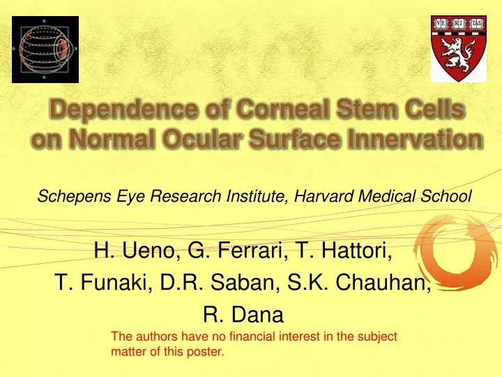
Dependence of Corneal Stem Cells on Normal Ocular Surface Innervation. H. Ueno, G. Ferrari, T. Hattori, T. Funaki, D.R. Saban , S.K. Chauhan , R. Dana. Schepens Eye Research Institute, Harvard Medical School. The authors have no financial interest in the subject matter of this poster.

E N D
Dependence of Corneal Stem Cellson Normal Ocular Surface Innervation H. Ueno, G. Ferrari, T. Hattori, T. Funaki, D.R. Saban, S.K. Chauhan, R. Dana Schepens Eye Research Institute, Harvard Medical School The authors have no financial interest in the subject matter of this poster.
Background Neurotrophickeratopathy (NK) is a degenerative disease of the cornea characterized by impaired healing. Corneal sensory nerve degeneration is responsible for corneal epithelial defects, stromal thinning, and perforation. Seitz B, Kruse FE. Ophthalmologe. 2005
Purpose • To identify the relationship between limbal stem cells and trigeminal innervation using a mouse model of corneal denervation. • Hypothesis: Neurotrophickeratopathy results in epithelial stem cell deficiency. • Approach: Analysis of stem cells in a mouse model of neurotrophickeratopathy.
Methods Trigeminal Stereotactic Electrolysis (TSE) no blink reflex 7days Cornea Ophthalmic nerve 1. Real Time-PCR ABCG2, p63, Hes1 2. Immunohistochemistry ABCG2, p63 3. Flow cytometry G. Ferrari et.al. preparation Side population
Expression of beta-III tubulin (neuronal marker) is decreased in the denervated cornea. Contralateral eye Denervated eye b-tubulinIII
Expression of stem cell markers (ABCG2 & p63) is decreased in the denervated cornea. Denervated Normal 2 ABCG2 DAPI ABCG2 DAPI p63 DAPI p63 DAPI
RNA expression levels for both stem cell markers ABCG2 and Hes1 are significantly decreased in denervated corneas. Relative Expression of mRNA Normal Expression of mRNA:1.0 p≦0.01 p≦ 0.01
Summary Corneal stem cells are significantly reduced in this murine model of neurotrophickeratopathy. Expression of stem cell markers (ABCG2, P63, Hes1) in denervated corneas is decreased at both the mRNA and protein levels.
Conclusion Our novel data clearly demonstrate that corneal nerves play a critical role in maintaining the corneal stem cells.
References 1. Touhami A, Grueterich M, Tseng SC. The role of NGF signalingin human limbal epithelium expanded by amniotic membrane Invest Ophthalmol Vis Sci. 2002 ;43:987-94. 2. Qi H, Chuang EY, Yoon KC, de Paiva CS, Shine HD, Jones DB, Pflugfelder SC, Li DQ. Patterned expression of neurotrophic factors and receptors in human limbal and corneal regions.Mol Vis. 2007 ;13:1934-41. 3. Qi H, Li DQ, Shine HD, Chen Z, Yoon KC, Jones DB, Pflugfelder SC. Nerve growth factor and its receptor TrkA serve as potential markers for human corneal epithelial progenitor cells. Exp Eye Res. 2008 ;86:34-40.