Download
1 / 74
740 likes | 1.21k Vues
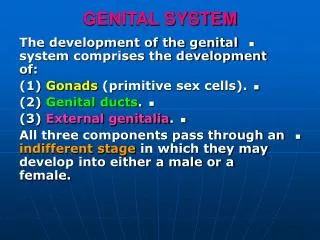
The Legendary Genius. MALE GENITAL SYSTEM. Development of Gonads. - During 5 th week: gonadal development occurs. - Until 7 th week: gonads are similar in both sexes. Development of Gonads. Gonads are derived from 3 sources :. Indifferent Gonads. Indifferent Gonads.

E N D
The Legendary Genius .. MALE GENITAL SYSTEM
Development of Gonads - During 5th week:gonadal development occurs . - Until 7th week:gonads are similar in both sexes .
Development of Gonads Gonads are derived from 3 sources :
Indifferent Gonads • the initial period of genital development is referred to as the indifferent stage of sexual development. • The initial stages of gonadal development occur during the fifth week ( 5 th )
A thickened area of mesothelium develops on the medial side of the mesonephros • Gonadal ridge:a bulgeon the medial side of mesonephros produced by: • Proliferation of mesothelium (cortex) • Proliferation (condensation) of mesenchyme (medulla)
Finger like epithelial cords or Gonadal cords soon grow into the underlying mesenchyme
The indifferent gonad now consists of: 1- an external cortex 2 - an internal medulla
Indifferent Gonads • In embryos with an XX sex chromosome complex, the cortex differentiates into an ovary and the medulla regresses • In embryos with an XY sex chromosome complex, the medulla differentiates into a testis and the cortex regresses • Except for vestigial remnants .
During folding of the embryo, the dorsal part of the yolk sac is incorporated into the embryo These large, spherical cells are visible early in the fourth week among the endodermal cells of the yolk sac near the allantois the primordial germ cells migrate along the dorsal mesentery of the hindgut to the gonadal ridges
Primordial Germ Cells • During the sixth week the primordial germ cells enter the underlying mesenchyme and are incorporated in the gonadal cords • The migration of the primordial germ cells is regulated by the genes : • 1- stella • 2-fragilis • 3- BMP-4. • If they fail to reach the ridges, the gonad remain indifferent or is absent…
Sex Determination • Chromosomal and genetic sex is determined at fertilization • It depends upon whether an X-bearing sperm or a Y-bearing sperm fertilizes the X-bearing ovum • The type of gonads develop is determined by the sex chromosome complex of the embryo (XX or XY)
Sex Determination • Before the seventh week, the gonads of the two sexes are identical in appearance called indifferent gonads • Development of the male phenotype requires a Y chromosome • The SRY gene for a testes-determining factor (TDF) has been localized in the sex-determining region of the Y chromosome ( on the short Arm of this chromosome ) • Two X chromosomes are required for the development of the female phenotype
Sex Determination • The Y chromosome has a testes-determining effect on the medulla of the indifferent gonad • The absence of a Y chromosome results in the formation of an ovary • Testosterone, produced by the fetal testes, determines the maleness • Primary female sexual differentiation in the fetus does NOT depend on hormones • It occurs even if the ovaries are absent • Expression of the Sox9 and Fgf9 genes is involved in the formation of the seminiferous cords.
Development of Testes • Embryos with a Y chromosome usually develop testes • The SRY gene for TDF on the short arm of the Y chromosome acts as the switch that directs development of indifferent gonad into testes
TDF induces the gonadal cords to condense and extend into the medulla of indifferent gonad, where they form rete testes
The connection of gonadal cords or seminiferous cords with the surface epithelium is lost as tunica albuginea develops
Development of Testes • The development of a dense tunica albuginea is the characteristic feature of testicular development in a fetus
The seminiferous cords develop into the seminiferous tubules, tubulirecti, and rete testis The enlarging testis separates from the degenerating mesonephros and becomes suspended by its own mesentery called mesorchium
The seminiferous tubules are separated by mesenchyme that gives rise to the interstitial cell of Leydig
Development of Testes • By the eighth week, these cells(Leydig) begin to secrete testosterone and androstenedione • These hormones induce masculine differentiation of the mesonephric ducts and external genitalia • Testosterone production is stimulated by HCG which reaches peak amounts during the 8- to 12-week period. • Fetal testes also produces a glycoprotein called antimullerian hormone (AMH) ormullerian inhibiting substance (MIS)
AMH suppresses development of the paramesonephric ducts • Seminiferous tubules remain solid until puberty
Development of Testes • The walls of seminiferous tubules are composed of two kinds of cells :
2 . Spermatogonia, primordial sperm cells derived from the primordial germ cells Sertoli cells, supporting cells derived from the surface epithelium on the testis
The rete testis becomes continuous with 15 to 20 mesonephric tubules that become efferent ductules - These ductules are connected with the mesonephric duct - It becomes the duct of the epididymis
Development of Genital Ducts • Both male and female embryos have two pairs of genital ducts • The mesonephric ducts (wolffian ducts) play an important role in the development of the male reproductive system • The paramesonephric ducts (mullerian ducts) have a leading role in the development of the female reproductive system • Till the end of sixth week, the genital system is in an indifferent state, when both pairs of genital ducts are present
A lateral outgrowth from the caudal end of each mesonephric duct gives rise to the seminal gland or vesicle The secretion from this pair of glands nourishes sperms Distal to the epididymis, the mesonephric duct acquires a thick investment of smooth muscle and becomes the ductus deferens The mesonephric duct between the duct of this gland and the urethra becomes the ejaculatory duct
Prostate • Multiple endodermal outgrowths arise from the prostatic part of the urethra • Grow into surrounding mesenchyme • The glandular epithelium of the prostate differentiates from these endodermal cells • The associated mesenchyme differentiates into the dense stroma and smooth muscle of the prostate
Bulbourethral Glands • pea-sized structures develop from paired outgrowths from the spongy part of the urethra . • The smooth muscle fibers and the stroma differentiate from the adjacent mesenchyme. • The secretions of these glands contribute to the semen.
Development of External Genitalia • Up to the seventh week of development the external genitalia are similar in both sexes • Distinguishing sexual characteristics begin to appear during the ninth week • External genitalia are not fully differentiated until the twelfth week
Early in the fourth week, proliferating mesenchyme produces a genital tubercle in both sexes at the cranial end of the cloacal membrane Labioscrotal swelling and urogenital folds soon develop on each side of the cloacal membrane The genital tubercle soon elongates to form a primordial phallus
Development of External Genitalia • When the urorectal septum fuses with the cloacal membrane, it divides it into : • A dorsal anal membrane • A ventral urogenital (Urethral) membrane
The urogenital membrane lies in the floor of a median cleft, the urogenital groove, which is bounded by urogenital folds
Development of Male External Genitalia • Masculization of the indifferent external genitalia is induced by testosterone
The phallus enlarges and elongates to become the penis The urogenital folds form the lateral walls of the urethral groove on the ventral surface of the penis to form the spongy urethra
The surface ectoderm fuses in the median plane of the penis, forming a penile raphe and enclosing the spongy urethra within the penis