Download
1 / 68
690 likes | 865 Vues
6th International Symposium on Translational Research in Oncology. October 11-14, 2007 Dublin, Ireland. This program is supported by educational grants from. 6th International Symposium on Translational Research in Oncology.

E N D
6th International Symposium on Translational Research in Oncology October 11-14, 2007Dublin, Ireland This program is supported by educational grants from
6th International Symposium on Translational Research in Oncology Dennis J. Slamon, MD, PhDChief, Division of Hematology/OncologyDavid Geffen School of Medicine at UCLALos Angeles, California John Crown, MD, MPHHead, Medical Oncology ResearchSt Vincent’s HospitalElm ParkDublin, Ireland Image crop is 3.5 x 5
Program Overview • Now in its sixth year, this annual symposium has a firmly established reputation as a premier meeting at which the world’s leading researchers gather to present and discuss new directions in oncology research with a focus on translating the most recent laboratory developments into improved clinical outcomes for cancer patients. Under the direction of John Crown, MD, MPH, and Dennis J. Slamon, MD, PhD, the program includes didactic presentations and interactive discussions. Faculty are carefully selected from among the researchers at the forefront of the translational work in the topic, whether from academia, government, or industry. The program encourages networking and interaction between the attendees and the renowned faculty members.
About These Slides • Users are encouraged to include these slides in their own presentations, but we ask that content and attribution not be changed. Users are asked to honor this intent. • These slides may not be published or posted online or used for any other commercial purpose without written permission from Clinical Care Options. • We are grateful to Gerry Melino, MD, PhD, the Chair of the Session, who aided in the preparation of this slideset. • We are also grateful to Donald W. Nicholson, PhD; Richard A. Knight, MD, PhD; Seamus J. Martin, PhD; Henning Walczak, PhD; and Gerry Melino, MD, PhD, who gave us permission to use a select group of their slides from the meeting to make this presentation possible. DisclaimerThe materials published on the Clinical Care Options Web site reflect the views of the authors, not those of Clinical Care Options, LLC, the CME providers, or the companies providing educational grants. The materials may discuss uses and dosages for therapeutic products that have not been approved by the United States Food and Drug Administration. A qualified healthcare professional should be consulted before using any therapeutic product discussed. Readers should verify all information and data before treating patients or using any therapies described in these materials.
Session III: Apoptosis and Programmed Cell Death Gerry Melino, MD, PhDProfessorMedical Research CouncilLeicester, United Kingdom
Core Components of the Apoptotic Pathway Death Signals Death Regulators • Bcl-2 family • IAPs and anti-IAPs • Usurpins • Phosphorylation Caspases Neurodegeneration Cancer Apoptotic “Victims”
Caspase Maturation and Activation p32 Dormant pro p17 p12 p20 p17 Active Substrate cleavage
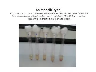
proCaspase-3 Levels in Human Colon Cancer Colonic Adenocarcinoma vs Adjacent Normal Mucosa 1 2 3 (Patient) 4 (Normal, Tumor) N T N T N T 6x 3 (Arbitrary Units/mg Protein) proCaspase-3 Content 2 p32 1 0 Normal Tumor (averaged data, n = 20)
Identification of an Intrinsic Regulatory “Safety Catch” Tripeptide A 179-181 Wt D28 C163 D175 D IPTG (min): 30 60 120 30 60 120 pro p17 p12 p32 DDD(179-181) ON p17 AAA OFF 1 2 3 4 5 6
The Safety Catch Modulates Vulnerability to Activator Proteases [Granzyme B][Caspase-9][proCaspase-3] C163A pro p17 p12 DDD(179-181) Safety catch ON AAA(179-181) Safety catch OFF 12 100 10 80 8 60 Caspase-9 Percentage Cleaved Percentage Cleaved 6 Granzyme B 40 4 proCaspase-3 20 2 0 0 0.001 0.01 0.1 1 10 100 1000 104 0.001 0.01 0.1 1 Protease (nM) proCaspase-3 (nM)
Summary: Caspase “Safety Catch” • Caspase-3 dormancy maintained by “safety catch” DDD regulatory tripeptide • Regulates cis autoactivation • Regulates trans activation by “initiator” caspases (GrznB, C9) • Caspase-3 autoactivation triggered by acidification • Destabilizes “safety catch” isoelectronic interactions • Relevant to in vivo activation mechanism • Therapeutics that antagonize “safety catch” DDD could preferentially sensitize or trigger apoptotic death
Nt AVPI--- IAP Antagonism During Apoptosis Mitochondrion SMAC/Diablo HtrA2/Omi ATP C APAF-1 C9 IAPs C3/C7 D A Apoptosis
Caspase Proteolysis Generates P1’-Ala Neo Termini Caspase Cleavage XIAP/hILP/hMIHA XIAP-AV (12mer) BIR1 BIR2 BIR3 RING Zn f SESD242 AVSSDRN AVPIAQK Smac/Diablo AVPSSPP hOmi/HtrA2 ATPFQEG hCaspase-9 p12 VEVD720 AAVTPEE Amyloid-ß Precursor Protein A APP-AA (31mer)
Functional Antagonism of IAPs in Cell-Free Extracts SMAC/Diablo APP XIAP 80 180 120 160 70 Smac-20 APP-AA XIAP-AV 100 140 60 APP-MG XIAP-MV 120 80 50 100 DEVDase Activity(% of Noninhibited Activity) DEVDase Activity (% of Noninhibited Activity) DEVDase Activity (% of Noninhibited Activity) 40 60 80 30 60 40 20 40 20 10 20 0 0 0 0 12.5 50 200 0 50 200 0 50 200 800 M M M
Summary: IAP Antagonism • IAP proteins are direct inhibitors of caspases • Ensure dormancy in healthy cells • Naturally antagonized by Smac and some caspase substrates after caspase proteolysis • IAP antagonism can be mediated by peptides and small molecules • Therapeutics that antagonize IAPs could preferentially sensitize or trigger apoptotic death
The p53 Family • The p53 family includes 3 genes, encoding for p53, p63, p73 proteins • The oldest protein is p63, whereas p53 is the most recent • P53 is involved in DNA damage repair • Each protein exists as different isoforms generated by distinct promoters (TA isoforms and N isoforms) or by alternative splicing (, , , etc, isoforms) • TA isoforms induce cell death (anticarcinogenic, acting as tumor suppressor) • N isoforms inhibit cell death (procarcinogenic, acting as oncogenes) • p73 and p63, like p53, are involved in DNA damage repair, inhibiting cell cycle progression, and inducing apoptosis • p73 is rarely mutated in cancer (0.5% mutation rate of p73 versus > 50% mutation rate of p53)
The p53 Family (cont’d) • p73 kills cells by regulating the transcription of genes crucial to the cell cycle (eg, p21) and to apoptosis (eg, bax, PUMA, CD95), thus affecting the sensitivity of cancer cells to chemotherapy • After 30 years of research on p53, clinically exploitable targets are mostly limited to its protein degradation regulation (inhibitors of the ubiquitin E3 ligase MDM2), and ARF (DNA methyltransferase inhibitors) pathways,
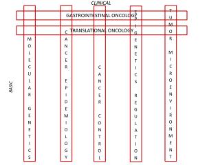
p63 Alterations DNA damage Replicative Stress DNA-PK Sensors PARP Ku70/Ku86 ATR ATM Signals chk2 HR NHEJ NER MMR BER c-abl chk1 p73 p53 mdm2 BRCA1 Effectors cdc25 rad50 NSB1 MRE11 14-3-3 cdc25 bax CD95 14-3-3 Noxa …. p21 gadd45 …. Cdc2 …. G1 S G2 Effects Apoptosis Cell Cyle Arrest DNA Repair Sayan et al. J Biol Chem. 2006;281:13566. Gong et al. Nature. 1999;399:806. Melino et al. J Biol Chem. 2004;279:8076. Bernassola et al. J Exp Med. 2004;199:1545. Gressner et al. EMBO J. 2005;24:2458. Mueller et al. Cell Death Differ. 2005;12:1564. Flinterman et al. J Biol Chem. 2005;280:5945. Vigano et al. EMBO J. 2006;25:5105. Rossi et al. PNAS. 2006;103:12753
p73 Promoter, Proteins, and Translational Targets Regulators of p73 promoter Signals Oncogenes: E2F1, c-Myc, Ras Viral proteins: E1A, Tax Drugs: retinoids Trascriptional inducers p73 interacting proteins Oncogenes: c-Myc, c-Abl, Hipk2, WT1 Viral proteins: E1A, E4orf6, Tax Cell cycle genes: p300, MM1, HMBG1 E3 ligases: MDM2, MDMX, Sumo1, PIAS1 Family members: p53, p63, p73 Interacting Proteins Regulatory Proteins p73 Posttranslational modifications Degradation pathways Evolution Transcriptional targets p73 trascriptional targets Effects Cell cycle genes: p21WAF1, gadd45, cyclin, kip2 Apoptosis: Bax, CD95, PUMA, perp, Noxa, p53AIP1 Signaling: 14-3-3, Pig13, RTP, ADTC, p53R2, IGFBP2, jun-B, IKKa family members: DeltaN-p73 Differentiation: AGP3, VEGF (neg), loricrin, involucrin, NCAM
Oncogenes DNA methyltransferase inhibitors ARF Ub E3 ligase inhibitors nutlin mdm2 DNA damage Activators PRIMA-1 p53 Cell cycle arrest Senescence Apoptosis
How Are p63/p73 Protein Levels Controlled? • Protein degradation of both p73 and p63 is regulated by the ubiquitin E3 ligase ITCH, a member of the HECT-containing E3s • In addition to p73 and p63, ITCH controls the degradation of c-jun, Notch, jun-B, and Flip, all involved in oncogenesis and apoptosis • The prediction is therefore that ITCH regulation affects carcinogenesis and/or chemosensitivity • Modulation of ITCH protein levels regulates chemosensitivity in vitro, suggesting that an inhibitor of ITCH could increase chemosensitivity
ITCH E3 Ligase Regulates p73 Stability TA PR DNA binding domain OD PR SAM TAp73 N C Phage display ITCH C2 WW HECTc WW N C Ca2+ dependent lipids interaction Protein-protein interacting domain Catalytic domain transferring ubiquitin to substrate Ubiquitin E3 ligase (NEDD4 family) 18H Mice natural Knockout (immune defects) Interacts with atrophin Degrades the transcription factor: Jun-B, c-Jun, Notch, Flip
504 ITCH Interacts With p63 Y504 Myc-ITCH -ITCH
Is ITCH E3 Activity Regulated? Summary • The function of ubiquitin E3 ligase ITCH is regulated by a physical interaction with a novel protein called N4BP1 • N4BP1 competes with ITCH substrates (p63, p73, c-Jun) by binding on the same region of ITCH, called WW2 • N4BP1 physiologically regulates ITCH and its substrates (p73, p63, c-Jun), thereby affecting cell death
a b c d N4BP1 Specifically Inhibits ITCH-Catalyzed Protein Substrates p73 p73 c-jun p53
UV-Induced Protein Stabilization of c-Jun Is Impaired in N4BP1-Deficient Cells +/+ -/- N4BP1 MEFs 1 8 24 1 8 24 hrs C UV C UV C UV C UV C UV C UV IB: -c-Jun c-Jun IB: -p21 IB: -actin A model for N4BP1 inhibition of ITCH substrate ubiquitylation UV Ub N4BP1 Ub N4BP1 Ub Ub Ub Ub N4BP1 N4BP1 Ub E2 E2 + c-Jun c-Jun Cell death HECT WW c-Jun HECT WW c-Jun c-Jun ITCH ITCH c-Jun
A Model for ITCH-Mediated Regulation of p73 Function N4BP1 p63 ITCH Ub, degradation p73 DNA damage Tumor cell Tumor cell ITCH ITCH N4BP1 Ub p73 Ub p73 p73 p73 ITCH p73 ITCH ITCH p73 p73 ITCH ITCH ITCH puma, noxa, CD95 Apoptosis
Can We Inhibit ITCH E3 Activity? • Small molecular inhibitors of ubiquitin E3 ligase ITCH have been identified • Potential ITCH inhibitors are currently under evaluation for their anticancer activity
Summary • p63/p73 are involved in DNA repair/cancer • p63/p73 regulate chemosensitivity in cancer • p63/p73 are ubiquitinated and degraded by ITCH • ITCH is regulated by N4BP1 • Low MW ITICH inhibitors are under development Perspective • ITCH is a candidate therapeutic target (to regulate p63/p73)
p53 • Is induced by DNA damaging agents • Determines cell cycle arrest (G1/S and G2/M ) • Induces apoptosis • Is frequently mutated (50%) or inactivated (20%) in all human cancers I II III IV V 100 200 300 393 TA DBD OD
The p53 proteins Genomic Structure Protein Structure TA PR DBD OD C N Transcription dependent Death Regulation Transcription Independent
Etoposide - + + - - + Z-VAD-fmk - 3 6 7 8 Caspase 50 37 25 25 Caspase 3 - + + + + p53 Is Cleaved by Caspases A B C - 20 50 100 200 400 nM Caspase 3 1 50 2 37 3 4 at D21 and D186 D186A D21A, D186A D21A WT WT p53 FL393 DO1 1801 C19 D E Caspase 3 - + - + - + - + 50 37 25 Panel A: p53 is processed to smaller fragments after etoposide treatment and this is abrogated by z-VAD. Panel B: Caspases 3, 6, 7, and 8 cleave p53 in vitro and, panel C, as little as 50nM caspase 3 is required. Panels D/E: by epitope mapping (panel D), the cleavage sites were mapped to D21 and D186 (panel E)
50 37 p32 p19 p17 p116 p85 p53 Is Cleaved During DNA Damage (*) A B (*) etoposide cisplatin, doxorubicin HCT116 U2OS Etoposide - + + - - - - - + + - - - - Cisplatin - - - + + - - - - - + + - - - - - - - + + - - - - - + + Doxorubicin - - + - + - + - - + - + - + Z-VAD-fmk P53 (C19) 37 P53 (DO1) 25 Caspase 3 PARP % apoptosis 6 37 65 14 32 10 4 44 51 9 6 5 12 7 1 2 3 4 5 6 7 8 9 10 11 12 13 14 lane
p116 p85 TRAIL - + + Z-VAD-fmk - - + 37 37 25 p53 Is Cleaved During Apoptosis (by TRAIL) A B SH-SY5Y HCT116 - 2 4 6 8 8 hours Etoposide Z-VAD-fmk - - - - - + P53 (C19) P53 (C19) 50 P53 (DO1) P53 (DO1) p32 Caspase 3 Caspase 3 p19 p17 PARP PARP
p53 + + + + Cisplatin - + - + 2 3 4 5 1 Fraction m m c/n c/n 50 Actin 37 37 PCNA 25 COXII 50 p53 37 25 Fragments Localize to Mitochondria B 1 21 186 393 A 75 CTD TA PRD DBD OD 50 FLp53 HA 37 V5-His p53(1-186) V5-His 25 p53(187-393) FLAG p53(22-186) V5-His p53(22-393) V5-His C D p53(1-393) p53(1-185) p53(187-393) p53(22-185) p53(22-393) Anti-p53 DAPI Merge Mito-RFP Fragments 1-186 and 22-186 localize to mitochondria by confocal (panel C) and biochemical fractionation (panel D)
p53 + + + + Cisplatin - + - + Fraction m m c/n c/n 50 Actin 37 37 PCNA 25 COXII 50 p53 37 25 Fragments Localize to Mitochondria (cont’d) A B (2) Biochemical fractionation D C
p53 Fragments Induce Apoptosis . . . Transcription Independent Both 1-186 and 22-186 induce apoptosis (panel A) which is transcription independent (panel B)
Mutant p53 Is Cleaved by Caspases . . . Contributing to Apoptosis 2 naturally occurring p53 mutants are also cleaved in a similar manner to the wt protein after cisplatin (panel B) and induce apoptosis (panel C), although their noncleavable mutants do not (circled)
What About the Other p53 Family Members (p73 and p63)? Alterations DNA Damage Replicative Stress DNA-PK Sensors PARP Ku70/Ku86 ATM ATR Signals chk2 HR NHEJ NER MMR BER c-abl chk1 p53 mdm2 p73/p63 BRCA1 Effectors cdc25 NSB1 rad50 MRE11 14-3-3 cdc25 bax CD95 14-3-3 Noxa …. p21 gadd45 …. Cdc2 …. G1 S G2 Effects Apoptosis Cell Cyle Arrest DNA Repair p73 and p63, members of the p53 family, cooperate with p53 in DNA damage response
p73 Is Cleaved During Apoptosis TRAIL-, etoposide-induced cleavage (reverted by caspase inhibitors) p73 is also cleaved by caspases after TRAIL and etoposide treatment
p73 Is Cleaved by Caspases in Vitro Caspase 3 is most effective All N-ter isoforms cleaved Caspase 8 also effective All C-ter isoforms cleaved
p63 Is Also a Caspase Target UV-induced cleavage In vitro cleavage by caspases Both N-ter isoforms cleaved D458 is the cleavage site p63 is cleaved by caspases in cells after UV-B (panel A) and in vitro (panel B). Both TA and DN isoforms are cleaved (panel C) and epitope mapping shows that D458 is the cleavage site (panels D/E)
TAp63 Cleavage Increases Transcriptional Activity Localization Transcriptional activity The N-terminal fragments of TA and DNp63 (TA-F and DN-F) are nuclear (panel A) and the TA fragment has enhanced transcriptional activity on 4 promoters compared to intact TAp63a
Endogenous p63 Is Cleaved During Apoptosis . . . by UV, staurosporine, cisplatin . . . mediated by caspases (not calpain inhibitors)
Summary • p53 is cleaved by caspases during apoptosis, contributing to cell death • Transcriptionally inactive natural mutants of p53 can be cleaved by caspases to produce transcriptionally inactive fragments • Can still induce apoptosis by depolarization of mitochondria • Other members of the p53 family, p63 and p73, are also susceptible to caspase cleavage • Caspase cleavage of p63a isoforms relieves the inhibitory effects of the C-terminal transactivation inhibitory domain, resulting in • Enhanced transcriptional activity by the TA isoform • Abrogating the transactivational inhibitory effects of the DNp63 isoform