Download
1 / 23
240 likes | 511 Vues
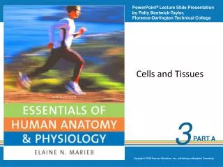
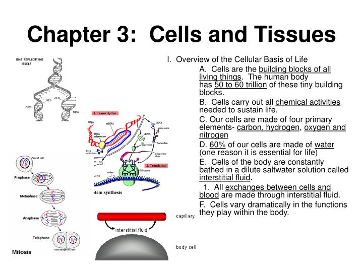
Chapter 3: Cells and Tissues. I. Overview of the Cellular Basis of Life A. Cells are the building blocks of all living things . The human body has 50 to 60 trillion of these tiny building blocks. B. Cells carry out all chemical activities needed to sustain life.

E N D
Chapter 3: Cells and Tissues I. Overview of the Cellular Basis of Life A. Cells are the building blocks of all living things. The human body has 50 to 60 trillion of these tiny building blocks. B. Cells carry out all chemical activities needed to sustain life. C. Our cells are made of four primary elements- carbon, hydrogen, oxygen and nitrogen D. 60% of our cells are made of water (one reason it is essential for life) E. Cells of the body are constantly bathed in a dilute saltwater solution called interstitial fluid. 1. All exchanges between cells and blood are made through interstitial fluid. F. Cells vary dramatically in the functions they play within the body.
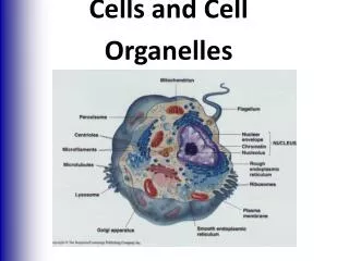
II. Anatomy of a Cell A. Cells are organized into three main regions 1. Nucleus a. contains DNA (genetic material) 2. Cytoplasm 3. Plasma Membrane a. barrier for cells contents b. double phospholipids bilayer c. Selectively permeable- regulates what enters leaves the cell Selective Transport Animations
d. Plasma membrane junctions; the meeting of two adjacent cell membranes. There are three types: • Tight junctions- fit together like a zipper forming an impermeable junction. These exist in cells lining the digestive tract to keep digestive enzymes and microorganisms from seeping into the bloodstream. • Desmosomes- anchoring junctions that prevent cell separation. These are abundant in tissues that are subjected to great mechnical stress, such as skin cells. • Gap junctions- the cells are connected by hollow cylinders (connexons) which allows chemical substances to pass between cells. These are abundant in heart muscle tissue where the movement of ions from cell to cell to synchronize the heart rhythm.
C. Cell Diversity 1. All cells share the same general structures; a cell membrane, a nucleus and cytoplasm. However, their function in the body is different. See examples below: Secretory vs. Absorptive Epithelial Picture
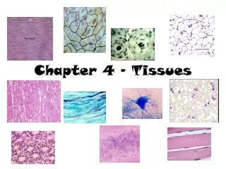
III. Body Tissues • Tissues are groups of cells with similar structure and function. • Four primary tissues types: • Epithelium (covering) • Connective (support) • Nervous Tissue (control) • Muscle (movement)
IV. Epithelium • Found in different areas of the body, such as body coverings, body linings, and glandular tissue. • Functions are for protection (skin), absorption (small intestine),filtration (kidneys), and secretion (glands). • Characteristics of epithelial tissue include: • Cells fit closely together • Tissue layer always has one free surface (the apical surface) that is exposed to the cavity of an internal organ or the body’s exterior. • The lower surface is bound by a basement membrane. • Avascular (these tissues have no blood supply of their own) • Regenerate well if nourished.
D. Classification of epithelium • Number of cell layers • simple- one layer • stratified- more than one layer • Shape of cells • squamous- flattened • cuboidal- cube shape • columnar-column- like
Types of Epithelium Thin for gas exchange between alveoli and capillaries Can secrete mucus and have cilia to help clean air Contain lots of golgi and ER to manufacture and secrete or absorb Many layers of cells to replace those lost when swallowing Adapted for secretion of mucus and ciliated to propel food Stretches as bladder fills with urine
V. Connective Tissue A. Found everywhere in the body, the most abundant and widely distributed tissue. B. Functions include: binding tissues together, support, and protection C. Characteristics of connective tissue: • Variations in blood supply- some tissue types are well vascularized (have good blood supply), while some have a poor blood supply (tendons and ligaments). Cartilage is avascular. • Extracellular matrix- the nonliving material that surrounds the tissue. (This is what makes connective tissue so different from other tissues.) • Matrix is composed of a ground substance (water,protein, and other molecules) and fibers (collagen,elastic, reticular). • The matrix allows connective tissue to act as a soft packing tissue around organs (adipose tissue), to bear weight, or withstand stretching and abrasion (bones, tendons and ligaments).
Connective Tissue D. Connective tissue types: • Bone (osseous) - composed of bone cells, hard matrix of calcium salts, large numbers of collagen fibers. • used to protect and support the body
Connective Tissue D. Connective tissue types: • Hyaline Cartilage- most common type of cartilage, composed of collagen and matrix • Entire fetal skeleton is hyaline cartilage, but by the time of birth, most cartilage is replaced by bone.
V. Connective Tissue D. Connective tissue types: • Elastic cartilage- provides elasticity • Example- supports the external ear • Fibrocartilage- highly compressible • Example- forms cushion-like discs between vertebrae
V. Connective Tissue D. Connective tissue types: • Dense connective tissue- main matrix element is collagen fibers which form strong rope-like structures, (the collagen producing cells are called fibroblasts) • Example- tendons (attach muscle to bone), ligaments (attach bone to bone)
V. Connective Tissue D. Connective tissue types: • Areolar connective tissue- most widely distributed connective tissue that serves as a kind of universal packing material between other tissues • contains all fiber types, • can soak up excess fluid (this is the tissue that swells causing edema) • universal packing tissue and “glue” that holds internal organs together
V. Connective Tissue D. Connective tissue types: • Adipose tissue- commonly called fat • matrix is Areolar tissue in which fat globules are predominate • these cells contain large deposits of lipids • functions to insulate the body, protect organs, and serves as a site of fuel storage
V. Connective Tissue D. Connective tissue types: • Reticular connective tissue-delicate network of interwoven fibers • forms internal network of lymphoid organs (lymph nodes, spleen, and bone marrow)
V. Connective Tissue D. Connective tissue types: • Blood- blood cells surrounded by a fluid matrix • fibers are visible during clotting • functions as the transport vehicle for materials
IV. Muscle Tissue • Functions to produce movement • Three types are: • Skeletal muscle- voluntary, striated • Smooth muscle – involuntary, surrounds organs • Cardiac muscle- involuntary, only in heart, striated • Intercalated disks are the junctions that allow heart cells to rapidly conduct electrical impulses through the heart.
VII. Nervous Tissue • Composed of neurons and nerve support cells • Functions to send and receive impulses to other areas of body • Located in nervous system structures such as the brain, spinal cord and nerves.
VIII. Tissue Repair (Wound Healing) A. Two types of tissue repair: 1. Tissue regeneration is the replacement of destroyed tissue by the same kind of cells 2. Fibrosis occurs when repair by dense fibrous connective tissue called scar tissue forms. Fibrosis occurs in cardiac and nervous tissues of the body.
B. The type of tissue repair depends on the type of tissue damageand the severity of the injury. C. Steps in Tissue repair 1. Capillaries become very permeable. 2. Clotting proteins and other substances seep into the injured area. 3. A clot is constructed to wall off the injured area (when the clot dries and hardens this forms the scab). 4. Formation of granulation tissue (delicate tissue that is made of new capillaries that grow into the damaged area).
a. this tissue also contains fibroblasts that synthesize collagen fibers that bride the gap 5. Surface epithelium regenerates; this covers an underlying layer of fibrosis (the scar). D. The regeneration of tissue 1. Tissues that regenerate easily: epithelial, fibrous connective, and bone 2. Tissues that regenerate poorly: skeletal muscle 3. Tissues that are replaced largely with scar tissue: cardiac and nervous tissue within the brain and spinal cord. Scar tissue lacks the normal flexibility of tissues which hinders the functioning. E. As we age there is a decrease in mass and viability of most tissues. The epithelia thin, the amount of collagen in the body declines which makes tissue repair less efficient, and nervous tissues begins to atrophy.