Download

1 / 22
220 likes | 426 Vues
TAQUICARDIA AURICULAR FOCAL EN PACIENTE TRASPLANTADO. Mª Fe Miguel Peña Unidad de Arritmias. INTRODUCCIÓN. Paciente de 51 años, trasplante cardiaco ortotópico (técnica biatrial) en 1996 por disfunción sistólica de VI de origen isquémico.

E N D
TAQUICARDIA AURICULAR FOCAL EN PACIENTE TRASPLANTADO Mª Fe Miguel Peña Unidad de Arritmias
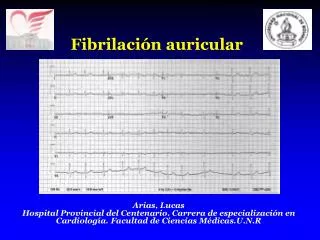
INTRODUCCIÓN • Paciente de 51 años, trasplante cardiaco ortotópico (técnica biatrial) en 1996 por disfunción sistólica de VI de origen isquémico. • Seguimientos sin incidencias, injerto normofuncionante, coronariografía sin lesiones en 2008. • Consulta por palpitaciones desde enero/2011. • ECG: ritmo sinusal a 95 lpm. QRS a +60º, BCRDHH. Alterna con taquicardia paroxística supraventricular a 190 lpm. • Ecocardiograma transtorácico: dilatación auricular derecha, función biventricular normal. • Biopsia endomiocárdica: sin evidencia de rechazo.
¿Cuál es el ritmo del trazado? • 1. Ritmo sinusal, bloqueo de rama izquierda • 2. Ritmo sinusal, bloqueo de rama derecha. • 3. Taquicardia supraventricular sostenida, bloqueo de rama izquierda • 4. Taquicardia supraventricular no sostenida, bloqueo de rama derecha
S T
¿Cuántas ondas P distintas se ven en el trazado? • 1. Una: sinusal. • 2. Dos: sinusal y ectópica • 3. Múltiples morfologías
INTERVALOS DE CONDUCCION AH HV
¿Dónde se encuentra el catéter de ablación? • 1. En la aurícula derecha. • 2. En la aurícula izquierda. • 3. En el haz de His • 4. En el ápex de ventrículo derecho
Halo con dipolos prox posteriores y distales anteriores (en Ritmo Sinusal)
Sinusal Taquicardia
Taquicardia con mec. focal: no ocupa la mayor parte del ciclo de la taquicardia
Posición de Halo. Proy. OAD Halo 9-10 Halo 19-20 Halo 1-2
Aplicación de RF Taquicardia Sinusal