Download
1 / 40
580 likes | 1.14k Vues
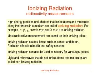
Ionizing Radiation in Medicine. There are 3 main uses of ionizing radiation in medicine:. Treatment Diagnosis Sterilisation. What is Cancer?. Cancers are growths of cells (cancerous tumors) which are out of control. As a result of this, they do not perform their intended function.

E N D
There are 3 main uses of ionizing radiation in medicine: • Treatment • Diagnosis • Sterilisation
What is Cancer? Cancers are growths of cells (cancerous tumors) which are out of control. As a result of this, they do not perform their intended function.
Treatment of Cancer • Cancerous tumors can be treated using the following main methods: • Chemotherapy (drugs). • Radiation therapy (radiotherapy and brachytherapy). • Surgery.
Factors Which Affect the Choice of Treatment for Cancer • The size of the tumor. • The position of the tumor. The choice of treatment depends on a number of factors including:
The Aims of Radiation Therapy The aim of radiation therapy is to cause damage to the cancerous cells while minimizing the risk to surrounding healthy tissue. The damage inflicted by radiation therapy causes the cancerous cells to stop reproducing and thus the tumor shrinks. Unfortunately, healthy cells can also be damaged by the radiation.
Why does the amount of radiation given to the patient have to be accurately calculated? The amount of radiation given to the patient has to be accurately calculated so that the damage is limited to the cancerous cells only.
Radiation Therapy Radiation therapy uses ionizing radiation to treat cancer i.e. to destroy cancerous cells. There are two techniques in radiation therapy that are used to treat cancer using ionizing radiation: • Radiotherapy • Brachytherapy
Radiotherapy Treatment Planning Every treatment using radiotherapy has to be rigorously planned. The planning process consists of three phases: • Planning • Simulation • Treatment
Radiotherapy Treatment PlanningPlanning • The cancerous tumor has to be located so that its size and position can be analyzed. • This information can be obtained from: • X-rays • CT scans • MRI scans • Ultrasound images
Radiotherapy Treatment PlanningSimulation Once the amount of radiation to be given has been accurately calculated, the patient then goes to the simulator to determine what settings are to be selected for the actual treatment using a linear accelerator. The settings are determined by taking a series of x-rays to make sure that the tumor is in the correct position ready to receive the ionizing radiation.
Radiotherapy Treatment PlanningTreatment Cancerous tumours can be treated using radiotherapy as follows: • Irradiation using high energy gamma rays. • Irradiation using high energy x-rays.
Radiotherapy TreatmentIrradiation Using High Energy Gamma Rays • Gamma rays are emitted from a cobalt-60 source – a radioactive form of cobalt. • The cobalt source is kept within a thick, heavy metal container. • This container has a slit in it to allow a narrow beam of gamma rays to emerge.
Radiotherapy TreatmentIrradiation Using High Energy X-rays • The x-rays are generated by a linear accelerator (linac). • The linac fires high energy electrons at a metal target and when the electrons strike the target, x-rays are produced. • The x-rays produced are shaped into a narrow beam by movable metal shutters.
Treatment of CancerRadiotherapy • The apparatus is arranged so that it can rotate around the couch on which the patient lies. • This allows the patient to receive radiation from different directions. • The diseased tissue receives radiation all of the time but the healthy tissue receives the minimum amount of radiation possible. • Treatments are given as a series of small doses because cancerous cells are killed more easily when they are dividing, and not all cells divide at the same time – this reduces some of the side effects which come with radiotherapy.
Radiation TherapyBrachytherapy • This involves placing implants in the form of seeds, wires or pellets directly into the tumor. • Such implants may be temporary or permanent depending on the implant and the tumor itself. • The benefit of such a method is that the tumor receives nearly all of the dose while healthy tissue hardly receives any.
Radiation TherapyBrachytherapy • Uterus • Cervix • Prostate • Intraocular • Skin • Thyroid • Bone Brachytherapy is used to treat the following cancers:
Tracers There are many uses of ionizing radiation based on the fact that it is easy to detect. In such applications, the radioactive material is used in the form of a tracer. In nuclear medicine, a tracer is a radioactive substance which is taken into the body either, as an injection, or as a drink. Such a substance is normally a gamma emitter which is detected and monitored. This gives an indication of any problems that may be present in body organs or tissues by how much, or how little, of the substance has been absorbed.
Nuclear MedicineTracers It is important to be able to study internal organs, or tissues, without the need for surgery. In such cases, radioactive tracers can be injected into the body so such studies can take place. The path of these tracers can be detected using a gamma camera because of their radioactivity. • A drug which is chosen for the particular organ that is being studied. • A radioactive substance which is a gamma emitter. Such tracers consist of two parts:
Factors Which Affect the Choice of Tracer • They will concentrate in the organ, or tissue, which is to be examined. • They will lose their radioactivity (short 1/2). • They emit gamma rays which will be detected outside the body. Such tracers are chosen so that:
Factors Which Affect the Choice of Tracer • Gamma rays are chosen since alpha and beta particles would be absorbed by tissues and not be detected outside the body. • Technitium-99m is most widely used because it has a half-life of 6 hours.
Why is a half-life of 6 hours important? • A shorter half-life would not allow sufficient measurements or images to be obtained. • A longer half-life would increase the amount of radiation the body organs or tissues receive. A half-life of 6 hours is important because:
The Gamma Camera The tracer is injected into the patient. The radiation emitted from the patient is detected using a gamma camera. A typical gamma camera is 40 cm in diameter – large enough to examine body tissues or specific organs. The gamma rays are given off in all directions but only the ones which travel towards the gamma camera will be detected.
The Gamma Camera A gamma camera consists of three main parts: • A collimator. • A detector. • Electronic systems. electronic systems detector collimator
The Gamma Camera • The collimator is usually made of lead and it contains thousands of tiny holes. • Only gamma rays which travel through the holes in the collimator will be detected. The Collimator
The Gamma Camera The Detector • The detector is a scintillation crystal and is usually made of Sodium Iodide with traces of Thallium added. • The detector is a scintillation crystal and it converts the gamma rays that reach it into light energy.
The Gamma Camera The Electronic Systems • The electronic systems detect the light energy received from the detector and converts it into electrical signals.
DiagnosisStatic Imaging • There is a time delay between injecting the tracer and the build-up of radiation in the organ. • Static studies are performed on the brain, bone or lungs scans.
DiagnosisDynamic Imaging • The amount of radioactive build-up is measured over time. • Dynamic studies are performed on the kidneys and heart.
Dynamic ImagingThe Renogram • To assess individual kidney and/or bladder function. • To detect urinary tract infections. • To detect and assess obstructed kidney(s). • To detect and assess vesico-ureteric reflux. • To assess kidney transplant(s). Renograms are dynamic images of the kidneys and they are performed for the following reasons:
Performing the Renogram • The tracer is injected into the patient. • The radioactive material is removed from the bloodstream by the kidneys. • Within a few minutes of the injection, the radiation is concentrated in the kidneys. • After 10 – 15 minutes, almost all of the radiation should be in the bladder. • The gamma camera takes readings every few seconds for 20 minutes.
DiagnosisThe Renogram • The computer adds up the radioactivity in each kidney and the bladder. • This can be shown as a graph of activity versus time – a time-activity curve.
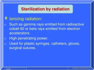
Sterilization • Radiation not only kills cells, it can also kill germs or bacteria. • Nowadays, medical instruments (e.g. syringes) are prepacked and then irradiation using an intense gamma ray source. • This kills any germs or bacteria but does not damage the syringe, nor make it radioactive.
Summary • Ionizing radiation is used in radiotherapy to treat cancer and to sterilise medical equipment because it destroys cells. • Radioactive tracers are used in nuclear medicine because the ionizing radiation it emits is easy to detect.
Summary • There are 3 main uses of ionizing radiation in medicine: treatment, diagnosis and sterilization. • Radiotherapy is used to treat cancers by irradiating them with ionizing radiation. • Radioactive tracers are used to diagnose and investigate several medical conditions. • Ionizing radiation is used to sterilize medical equipment as it kills germs and/or bacteria.
Careers • Oncologist. • Medical Physicist. • Radiographer. • Radiation Protection. • Environmental Protection. • Dosimetrist. • Research.