Download
1 / 3
30 likes | 215 Vues
Biomedical Research. Reactive Crystallization of Calcium Hydroxyapatite. Alison Mello Department of Chemistry and Biochemistry University of Massachusetts – Dartmouth . Dapeng Li Department of Materials and Textiles University of Massachusetts – Dartmouth . Chen-Lu Yang

E N D
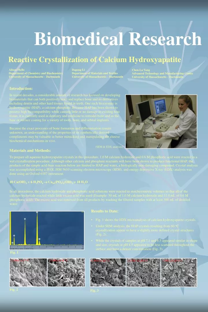
Biomedical Research Reactive Crystallization of Calcium Hydroxyapatite Alison Mello Department of Chemistry and Biochemistry University of Massachusetts – Dartmouth Dapeng Li Department of Materials and Textiles University of Massachusetts – Dartmouth Chen-Lu Yang Advanced Technology and Manufacturing Center University of Massachusetts – Dartmouth Introduction: In recent decades, a considerable amount of research has focused on developing biomaterials that can both positively react and replace bone and its derivatives including dentin and other hard tissues found in teeth. One such bioceramic is hydroxyapatite (HAP), a calcium phosphate. Because HAP has been shown to possess high biocompatibility while causing little or no damage to existing host tissue, it is currently used in dentistry and medicine to remodel bone and as the base or surface coating for a variety of tooth, bone, and orbital implants. Because the exact processes of bone formation and differentiation remain unknown, an understanding of the properties of its synthetically derived compliments may be valuable in better mimicking and manipulating the elusive biochemical mechanisms in vivo. (SEM & EDX analysis) Materials and Methods: To prepare all aqueous hydroxyapatite crystals in this procedure, 1.0 M calcium hydroxide and 0.6 M phosphoric acid were reacted in a wet crystallization procedure. Although other calcium and phosphate reactants will have been shown to produce functional HAP, the products of the simple acid-base reaction below are limited to HAP and water, a biologically non-damaging compound. Crystal analysis was accomplished using a JEOL JSM 5610 scanning electron microscope (SEM), and energy depressive X-ray (EDX) analysis was done using an Oxford 6587 instrument. 10 Ca(OH)2 + 6 H3PO4 Ca10(PO4)6(OH)2 + 18 H2O In all procedures, the calcium hydroxide and phosphoric acid solutions were reacted in stoicheiometric volumes so that all of the calcium hydroxide reacted while little excess acid was used (Example: 50 mL of 1.0 M calcium hydroxide and 51.0 mL of 0.6 M phosphoric acid). The excess acid was removed from all products by washing the filtered samples with at least 300 mL of distilled water. • Results to Date: • Fig. 1 shows the EDX microanalysis of calcium hydroxyapatite crystals. • Under SEM analysis, the HAP crystals resulting from 60 oC crystallization appear to have a slightly more defined crystal structures (Fig. 2). • While the crystals of samples at pH 7.1 and 8.5 appeared similar in shape and size, crystals at pH 8.5 appeared to be less scattered throughout the surface and have a denser concentration (Fig. 3). Fig. 1 (1,000x) at 27 oC (1,000x) at 60 oC (1,400x) at pH 7.1 (1,200x) at pH 8.5 (500x) at pH 11.5 Fig. 2 Fig. 3
@ ATMC • Results (continued): • SEM imaging of the samples revealed that crystals of the 2.0 bar samples possessed elongated oval-like crystals that were uniform in size and shape (Fig. 4). • Fig. 5. shows the consequences of additional heating of dry crystal. Qualitative analysis of the samples revealed that the additionally heated samples were all harder than the control sample. Furthermore, the samples heated for 24 hours were much harder than their 12 hour, same-temperature counterparts, and the samples heated at 150 oC were both harder than the crystals heated at 100 oC. SEM imaging revealed the overall size and shape of the crystals in all the over dried samples were all similar; they all possessed surfaces with irregular and non-uniformly shaped or sized crystal structures. Novel crystals seen in these samples were interspersed within the same viewing field. Structures were needlelike, fernlike, blossom-shaped, and starburst-patterned. (1,000x) at 1.01 bar (1,100x) at 2.0 bar (2,000x) at 2.0 bar (3,000x) at 2.0 bar Fig. 4 Fig. 5 (110x) varied crystal structures (350x) front fernlike pattern (300x) starburst patterns (800x) fernlike crystal (950x) flower pattern (2,300x) flower pattern center (300x) starburst pattern (1000x) flower pattern inverted position (9,000x) front needlelike crystal (430x) fernlike, needlelike, and irregular crystals (1,000x) needlelike crystals (700x) needlelike crystals Dye-sensitized Antibody-nanoparticle Conjugates for Rapid Detection of Residual Antibiotics Bin Wu Phosphorex, Inc. Fall River, MA Yue Huo Department of Materials and Textiles University of Massachusetts – Dartmouth Chen-Lu Yang Advanced Technology and Manufacturing Center University of Massachusetts – Dartmouth Introduction: Immunochromatographic strip (ICS) tests have been applied to more than 100 infectious diseases and more than 60 other chemical analytes such as hCG, RF, CRP, ASO, FDP, and fecal occult blood. The popular urine tests are AMP for amphetamine, BAR for barbiturates, BZO for benzodiazepines, COC for cocaine, MET for methamphetamine, MOP for morphine, MTD for methadone, OPI for Opiate, PCP for phencyclidine, TCA for tricyclic, and THC for marijuana. Due to recent reports on misuses and over-uses of antibiotics by doctors and patients, a need on urine test for antibiotics is on the horizon. Excitation Antibiotics Nanoparticle Emission Antibody Fluorescent Dye Fig. 1 The basic principles of dye-sensitized antibody-nanoparticle conjugation.
University of Massachusetts Dartmouth Chapter of Sigma Xi, the Scientific Research Society 14th Annual UMass Dartmouth Research Exhibition • Basic Principles: • By attaching antibody molecules to latex nanoparticles, the conjugates are prone to target antibiotic molecules through the antibody-antigen interaction mechanism (Fig. 1). • The signal from a single antibiotic molecule can be greatly enhanced by hundreds of fluorescent dye molecules encapsulated in the antibody-nanoparticle conjugate. • With the enhancement of the dye molecules on the conjugates, the detection limit of a fluorometer or spectrofluorometer can reach 5 - 50 ppb. • When properly designed, the latex nanoparticles will allow the detection of multiple antibiotics on one testing strip. • Experimental: • Buffer preparation • Synthesis of color nanoparticles • Synthesis of fluorescent nanoparticles • Conjugating nanoparticles to antibiotic antibodies • Confirmation of conjugation Table 1 • Results: • Table 1 shows the characteristics of color and fluorescent nanoparticles. • Figure 2 shows the nanoparticles of various colors. • Figure 3 shows the Zeta-Plus particle size distribution of blue nanoparticles, mean size: 190 nm, polydispersity 0.005. Fig. 3 • Table 2 shows the particle size of antibody-nanoparticle conjugates before and after addition of antigen measured by light scattering. • Figure 4 shows a fluorescent spectrum of the fluorescent nanoparticles prepared in this study (Entry 4 in Table 1). Rhodamine was the dye used in the preparation of the nanoparticles. The excitation wavelength was set at 540 nm, the emission peak is at 575 nm. • Figure 5 shows the absorbance of anti-Penicillin (200 ml/ml) at 280 nm. It clearly shows the 280 nm absorption of the antibody. • Figure 6 shows the UV spectrum of the antibody-nanoparticle conjugate suspension in DI water. Fig. 2 Table 2 Anti-Penicillin Control Anti-P Nanoparticle Conjugates ABS ABS Wavelength (nm) Wavelength (nm) Fig. 4 Fig. 5 Fig. 6