Download
1 / 28
320 likes | 739 Vues
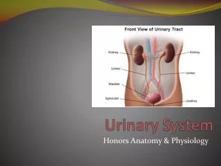
Urinary System. Chapter 25. urinary bladder. Urinary System. Two kidneys. Two ureters. Urethra. Functions of the Kidneys. Filters blood plasma, eliminates waste, returns useful chemicals to blood Regulates blood volume and pressure Secretes aldosterone controls BP, electrolyte balance

E N D
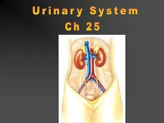
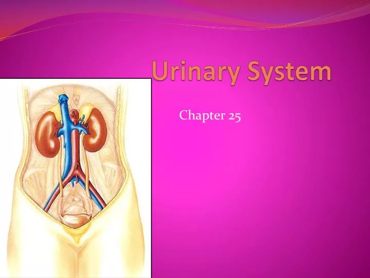
Urinary System Chapter 25
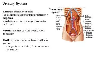
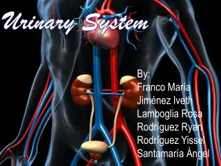
urinary bladder Urinary System • Two kidneys • Two ureters • Urethra
Functions of the Kidneys • Filters blood plasma, eliminates waste, returns useful chemicals to blood • Regulates blood volume and pressure • Secretes aldosterone • controls BP, electrolyte balance • Secretes erythropoietin, controls RBC count • Regulates acid base balance • Detoxifies free radicals and drugs • Remove nitrogenous wastes
Nitrogenous Wastes • Urea • proteinsamino acids NH2 removed forms ammonia, liver converts to urea • Uric acid • nucleic acid catabolism • Creatinine • creatinine phosphate catabolism • Renal failure • azotemia: nitrogenous wastes in blood • uremia: toxic effects as wastes accumulate
Kidneys as Filters • Diuretic- loose water; coffee, alcohol • Antidiuretic- retain water; ADH • Aldosterone- sodium & water reabsorption, and K+excretion
Anatomy of Kidney • Position, weight and size • Level of T12 to L3 • about 160 g each • about size of a bar of soap (12x6x3 cm) • Shape • lateral surface - convex; medial - concave • CT coverings • renal fascia: binds to abdominal wall • adipose capsule: cushions kidney • renal capsule: encloses kidney like cellophane wrap
renal pyramids renal pelvis renal cortex renal capsule ureter renal medulla Kidney Anatomy
Path of Blood Through Kidney • Renal artery interlobararteries(up renal columns, between lobes) arcuatearteries (over pyramids) interlobulararteries (up into cortex) afferentarterioles glomerulus (cluster of capillaries) efferentarterioles (near medulla vasa recta) peritubular capillaries interlobular veins arcuate veins interlobar veins • Renal vein
Proximal Convoluted Tubule Reabsorbs: water, glucose, amino acids, and sodium. • 65% of Na+ is reabsorbed • 65% of H2O is reabsorbed • 90% of filtered bicarbonate (HCO3-) • 50% of Cl- and K+
Composition and Properties of Urine • Appearance • almost colorless to deep amber; yellow color due to urochrome, from breakdown of hemoglobin (RBC’s) • Odor - bacteria degrade urea to ammonia • pH - range: 4.5 - 8.2, usually 6.0 • Chemical composition: 95% water, 5% solutes • urea, NaCl, KCl, creatinine, uric acid
Urine Volume • Normal volume - 1 to 2 L/day • Polyuria > 2L/day • Oliguria < 500 mL/day • Anuria - 0 to 100 mL
Urine Storage and Elimination • Ureters • from renal pelvis passes dorsal to bladder and enters it from below, about 25 cm long • 3 layers • adventitia - CT • muscularis - 2 layers of smooth muscle • urine enters, it stretches and contracts in peristaltic wave • mucosa - transitional epithelium • lumen very narrow, easily obstructed
Urinary Bladder • Located in pelvic cavity, posterior to pubic symphysis • 3 layers • parietal peritoneum • muscularis: detrusor muscle, 3 layers of smooth muscle • mucosa: transitional epithelium • trigone: openings of ureters and urethra, triangular • rugae: relaxed bladder wrinkled, highly distensible • capacity: moderately full - 500 ml, max. - 800 ml
Sphincter Muscles on Bladder • Internal urethral sphincter: • Smooth muscle • Involuntary control • More superiorly located • External Urethral sphincter: • Skeletal muscle • Voluntary control • Posteriorly located
ureters internal sphincters external sphincters urethra Urinary Bladder
Female Urethra • 3 to 4 cm long • External urethral orifice • between vaginal orifice and clitoris • Internal urethral sphincter • detrusor muscle thickened, smooth muscle, involuntary control • External urethral sphincter • skeletal muscle, voluntary control
Male Bladder and Urethra • 18 cm long • Internal urethral sphincter • External urethral sphincter • 3 regions • prostatic urethra • Receives semen • membranous urethra • Passes through pelvic cavity • penile urethra
Voiding Urine - Micturition • Micturition reflex 1) 200 ml urine in bladder, stretch receptors send signal to spinal cord (S2, S3) 2) parasympathetic reflex arc from spinal cord, stimulates contraction of detrusor muscle 3) relaxation of internal urethral sphincter 4) this reflex predominates in infants
Voluntary Control of Micturition 5) micturition center in pons receives stretch signals and integrates cortical input (voluntary control) 6) sends signal for stimulation of detrussor and relaxes internal urethral sphincter 7) to delay urination impulses sent through pudendal nerve to external urethral sphincter keep it contracted until you wish to urinate 8) valsalva maneuver • aids in expulsion of urine by pressure on bladder • can also activate micturition reflex voluntarily