Download
1 / 27
450 likes | 959 Vues
基因工程與原理. GENE CLONING AND DNA ANALYSIS. Chapter 3 Purification of DNA from Living Cells. 3.1 Preparation of total cell DNA 3.1.1 Growing and harvesting a bacterial culture 3.1.2 Preparation of a cell extract 3.1.3 Purification of DNA from a cell extract

E N D
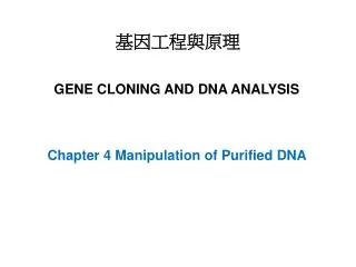
基因工程與原理 GENE CLONING AND DNA ANALYSIS Chapter 3 Purification of DNA from Living Cells
3.1 Preparation of total cell DNA 3.1.1 Growing and harvesting a bacterial culture 3.1.2 Preparation of a cell extract 3.1.3 Purification of DNA from a cell extract Removing contaminants by organic extraction and enzyme digestion Using ion-exchange chromatography to purify DNA from a cell extract 3.1.4 Concentration of DNA samples 3.1.5 Measurement of DNA concentration 3.1.6 Other methods for the preparation of total cell DNA 3.2 Preparation of plasmid DNA 3.2.1 Separation on the basis of size 3.2.2 Separation on the basis of conformation Alkaline denaturation Ethidium bromide–caesium chloride density gradient centrifugation 3.2.3 Plasmid amplification 3.3 Preparation of bacteriophage DNA 3.3.1 Growth of cultures to obtain a high λ titer 3.3.2 Preparation of non-lysogenic λ phages 3.3.3 Collection of phages from an infected culture 3.3.4 Purification of DNA from 2 phage particles 3.3.5 Purification of M13 DNA causes few problems
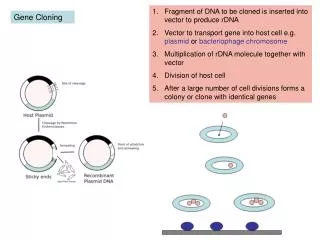
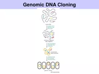
3.1 Preparation of total cell DNA Figure 3.1 The basic steps in preparation of total cell DNA from a culture of bacteria.
3.1.1 Growing and harvesting a bacterial culture Defined medium Complex or undefined Tryptone 蛋白胴: digestion of casein by the protease trypsin; amino acids and small peptides Yeast extract: nitrogen, sugar, inorganic and organic nutrients
Figure 3.2 Estimation of bacterial cell number by measurement of optical density (OD). (a) A sample of the culture is placed in a glass cuvette and light with a wavelength of 600 nm shone through. The amount of light that passes through the culture is measured and the OD (also called the absorbance) calculated as: Intensity of transmitted light 1 OD unit = log10 Intensity of incident light (b) The cell number corresponding to the OD reading is calculated from a calibration curve. This curve is plotted from the OD values of a series of cultures of known cell density. For E. coli,1 OD unit = 0.8 × 10⁹ cells/ml.
Figure 3.3 Harvesting bacteria by centrifugation.
3.1.2 Preparation of a cell extract Lysozyme: digest the cell wall EDTA: remove magnesium, inhibit DNase SDS: disrupt the cell membrane Figure 3.4 Preparation of a cell extract. (a) Cell lysis. (b) Centrifugation of the cell extract to remove insoluble debris.
Ion-exchange chromatography Figure 3.5 Two approaches to DNA purification. (a) Treating the mixture with reagents which degrade the contaminants, leaving a pure solution of DNA. (b) Separating the mixture into different fractions, one of which is pure DNA.
or phenol and chloroform (1:1) Protease (pronase or proteinase K) before phenol extraction Ribonuclease degrades RNA Figure 3.6 Removal of protein contaminants by phenol extraction.
DNA and RNA are negatively charged Figure 3.7 DNA purification by ion-exchange chromatography. (a) Attachment of DNA to ion-exchange particles. (b) DNA is purified by column chromatography. The solutions passing through the column can be collected by gravity flow or by the spin column method, in which the column is placed in a low-speed centrifuge.
3.1.4 Concentration of DNA samples Ethanol precipitation Monovalent cation (Na+) -20 ℃ or less Absolute ethanol Figure 3.8 Collecting DNA by ethanol precipitation. (a) Absolute ethanol is layered on top of a concentrated solution of DNA. Fibers of DNA can be withdrawn with a glass rod. (b) For less concentrated solutions ethanol is added (at a ratio of 2.5 volumes of absolute ethanol to 1 volume of DNA solution) and precipitated DNA collected by centrifugation.
3.1.5 Measurement of DNA concentration Ultraviolet (UV) absorbance spectrophotometry 1 OD (A260)= 50 mg/ ml dsDNA A260/ A280 ~ 1.8
3.1.6 Other methods for the preparation of total cell DNA Cetryltrimethylammonium bromide (CTAB) Figure 3.9 The CTAB method for purification of plant DNA.
Figure 3.10 DNA purification by the guanidinium thiocyanate and silica method. (a) In the presence of guanidinium thiocyanate, DNA binds to silica particles. (b) DNA is purified by column chromatography.
3.2 Preparation of plasmid DNA 3.2.1 Separation on the basis of size Figure 3.11 Preparation of a cleared lysate.
3.2.2 Separation on the basis of conformation Covalently closed-circular (ccc) Relaxed Figure 3.12 Two conformations of circular double-stranded DNA: (a) supercoiled—both strands are intact; (b) open-circular—one or both strands are nicked.
Non-supercoiled DNA is denatured Figure 3.13 Plasmid purification by the alkaline denaturation method.
Figure 3.14 Caesium chloride density gradient centrifugation. (a) A CsCl density gradient produced by high speed centrifugation. (b) Separation of protein, DNA, and RNA in a density gradient.
Figure 3.15 Partial unwinding of the DNA double helix by EtBr intercalation between adjacent base pairs. The normal DNA molecule shown on the left is partially unwound by taking up four EtBr molecules, resulting in the “stretched伸長” structure on the right.
Figure 3.16 Purification of plasmid DNA by EtBr–CsCl density gradient centrifugation.
Some multicopy plasmids can replicate in the absence of protein synthesis Figure 3.17 Plasmid amplification.
3.3 Preparation of bacteriophage DNA The maxinum titer for λ is 1010 /ml; ~ 500 ng DNA Figure 3.18 Preparation of a phage suspension from an infected culture of bacteria.
3.3.1 Growth of cultures to obtain a high λ titer Temperature-sensitive (ts) Figure 3.19 Induction of a λ cIts lysogen by transferring from 30°C to 42°C.
3.3.2 Preparation of non-lysogenic λ phages Deletions of the cI and other genes. Figure 3.20 Achieving the right balance between culture age and inoculum size when preparing a sample of a non-lysogenic phage.
3.3.3 Collection of phages from an infected culture Figure 3.21 Collection of phage particles by polyethylene glycol (PEG) precipitation.
3.3.4 Purification of DNA from λ phage particles Figure 3.22 Purification of λ phage particles by CsCl density gradient centrifugation.
3.3.5 Purification of M13 DNA causes few problems 1012 M13/ml Figure 3.23 Preparation of single-stranded M13 DNA from an infected culture of bacteria.