Download
1 / 19
230 likes | 1.02k Vues
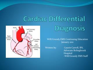
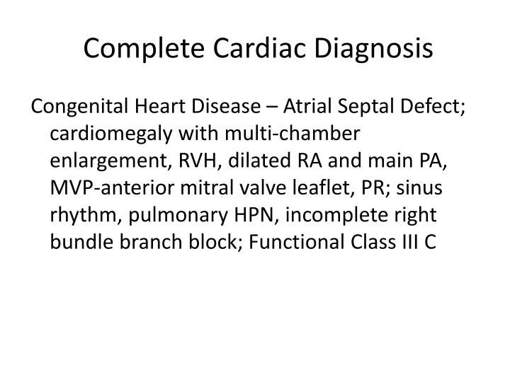
Complete Cardiac Diagnosis. Congenital Heart Disease – Atrial Septal Defect; cardiomegaly with multi-chamber enlargement, RVH, dilated RA and main PA, MVP-anterior mitral valve leaflet, PR; sinus rhythm, pulmonary HPN, incomplete right bundle branch block; Functional Class III C.

E N D
Complete Cardiac Diagnosis Congenital Heart Disease – Atrial Septal Defect; cardiomegaly with multi-chamber enlargement, RVH, dilated RA and main PA, MVP-anterior mitral valve leaflet, PR; sinus rhythm, pulmonary HPN, incomplete right bundle branch block; Functional Class III C
Patient ASD VSD PDA RHD Hx “heart disease” in childhood frequent URTI easy fatigability occasional chest pain Prolonged symptom-free period Palpitations Fatigue Dyspnea on exertion Orthopnea Frequent respiratory infections Symptoms of right ventricular failure most children with small defects remain asymptoma-tic In patients with Eisenmenger syndrome, symptoms in adult life consist of exertional dyspnea, syncope and hemoptysis R-L shunt leads to cyanosis, clubbing, and erythorocy-tosis premature birth, perinatal distress, or perinatal hypoxia may be present (+) upper respiratory tract infection Mitral stenosis -dyspnea and cough on exertion -orthopnea and PND -hemoptysis Mitral regurgitation -dyspnea and cough on exertion -orthopnea and PND -ankle edema
Patient ASD VSD PDA RHD P.E. Hyposthenic, narrow AP chest diameter Normal JVP and CAP Left lower sternal lift Normal S1 ff. by gr. 3/6 crescendo-decrescendo murmur S2 wide with fixed splitting Multiple clicks at apex Prominent RV impulse S1 normal or split, with accentuation of TV closure sound Wide & fixed splitting of the S2 Systolic ejection murmur (heard in pulmonic area) Diastolic rumble across the tricuspid valve Neck vein distention Ascites Edema loud, harsh, or blowing holosystolic murmur is heard best over the lower LSB in the 3rd or 4th ICS displaced cardiac apex with a similar holosystolic murmur apical diastolic rumble and third heart sound (S3) precordial activity is increased apical impulse is laterally displaced S1 normal, S2 typically obscured by murmur Continuous machinery-like murmur Bounding peripheral pulses Mitral stenosis diastolic thrill at the apex S1 and P2 are accentuated S2 is split or fixed OS of the mitral valve on expiration (+) carvallo's sign Mitral regurgitation (+) systolic thrill at the apex holosystolic murmur -(+) S4
Patient ASD VSD PDA RHD ECG Normal sinus rhythm RVH Incomplete RBBB Diffuse ST-T changes Ostium secundum defects – incomplete right bundle block & right axis deviation Ostium primum defects – left anterior hemiblock & left axis deviation May be normal With larger defects there are various degrees of right axis deviation associated with right ventricular Enlargement LVH with a larger PDA LVH
Patient ASD VSD PDA RHD CXR Cardiomegaly with multi- chamber enlargement and pulmonary congestion Shunt vascularity (inc. pulm. vascular markings) Right ventricular enlargement Enlargement of the pulmonary artery segment in P-A view Right Ventricular enlargement Increased pulmonary vascular markings LAE LVE PAE PVE Mitral stenosis -(+) kerley B lines -concentric hypertrophy of left atrium -cephalization -prominent main pulmonary artery and branches -constriction of arteries in the middle and peripheral lung zones Mitral regurgitation -eccentric hypertrophy of the left atrium -LA and LV enlargement -equalization, cephalization -mitral annulus calcification
Patient ASD VSD PDA RHD Echo ASD, ostium secundum type Markedly dilated right ventricle with adequate wall motion and contractility with evidence of RV pressure and volume overload Dilated RA w/o thrombus Dilated MPA Severe TR PR Mod. Pul. HPN Reverse E/A across mitral valve Enlargement of RV Negative-contrast image at the site of defect (saline injection) Doppler – abnormal pressure of left-to-right blood flow across the septum color flow can show the shunting of blood from the left ventricle to the right VSD's can result in a shunt from below the tricuspid valve to below the pulmonary valve. LAE Continuous flow from the aorta into the main pulmonary artery Mitral stenosis -mitral orifice <4cm -concentric hypertrophy of left atrium -annular calcifications Mitral regurgitation -eccentric hypertrophy of the left atrium -LA enlargement -hyperdynamic LV -annular calcifications/ LV dyskinesis -ruptured chordae tendineae
Differentiate Pulmonary arterial hypertension from pulmonary venous congestion
Pulmonary Arterial Hypertension • Causes • Primary/ Idiopathic • Genetic • Secondary • Cardiac • Pulmonary • hypoxic vasoconstriction • decreased area of the pulmonary vascular bed • volume/pressure overload
Secondary Pulmonary Arterial Hypertension • Hypoxic Vasoconstriction • COPD and obstructive sleep apnea • Due to down regulation of endothelial nitric oxide synthetase • Decreased Area of Pulmonary Bed • Occurs when loss of vessels exceed 60% of the total pulmonary vasculature • Occurs in patients with collagen vascular disease like CREST and scleroderma. And those with chronic emboli
Volume/ Pressure Overload • Seen in patients with left to right intracardiac shunts • May passively occur in patients with left atrial hypertension and left ventricular dysfunction, mitral valve disease and hose with aortic stenosis
Pulmonary Arterial Hypertension • Chest radiograph • Classic finding is enlargement of central of pulmonary arteries, attenuation of peripheral vessels and oligemic lung fields • Findings of RV and RA dilatation are possible
Pulmonary Venous Hypertension • Secondary to increased resistance to pulmonary venous drainage • Associated with diastolic dysfunction of the LV and valvular dysfunction • Features • Capillary congestion • Focal alveolar edema • Dilatation of interstitial lymphatics
mild cardiomegaly • normal pulmonary arterial markings • pulmonary venous congestion • fluid within the horizontal fissure • prominent Kerley B lines (indicative of lymphatic engorgement) Lateral chest film show marked venous congestion with fluid visible in both the horizontal and oblique fissures