Download
1 / 33
350 likes | 557 Vues
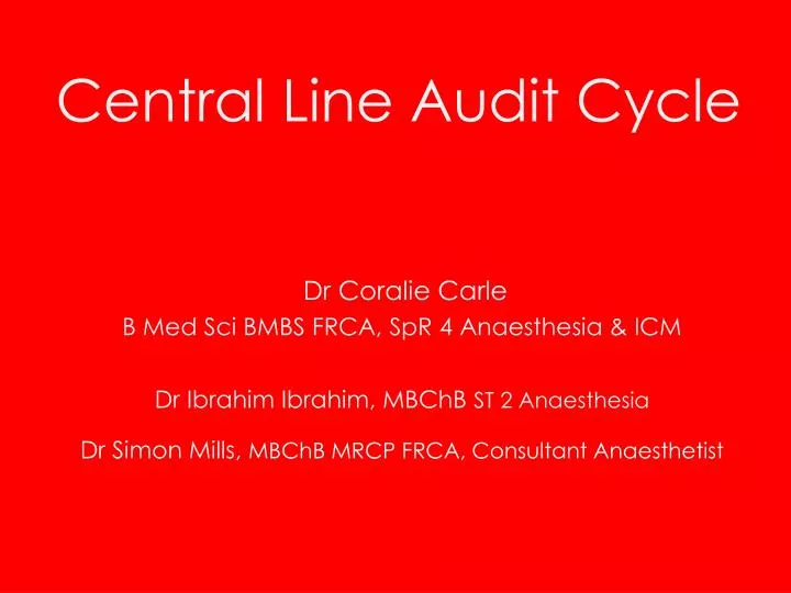
Central Line Audit Cycle. Dr Coralie Carle B Med Sci BMBS FRCA, SpR 4 Anaesthesia & ICM Dr Ibrahim Ibrahim, MBChB ST 2 Anaesthesia Dr Simon Mills, MBChB MRCP FRCA, Consultant Anaesthetist. Outline. trigger for audit background service evaluation intervention re-audit future plans.

E N D
Central Line Audit Cycle Dr Coralie Carle B Med Sci BMBS FRCA, SpR 4 Anaesthesia & ICM Dr Ibrahim Ibrahim, MBChB ST 2 Anaesthesia Dr Simon Mills, MBChB MRCP FRCA, Consultant Anaesthetist
Outline • trigger for audit • background • service evaluation • intervention • re-audit • future plans
Patient in PACU… • 37 year old male • post-op exploration of bleeding pseudoaneurysm / ileofemoral bypass • PMH • IVDU • Hep C +ve • PE (patient consent for presentation obtained)
…in extremis • acutely SOB in PACU • ABC approach with simultaneous consideration of diagnoses • pneumothorax • PE • transfusion reaction • air embolism
CVC inspection • 3-way stopcock aligned so it was potentially open to the atmosphere • partially loose (cross threaded) red replacement cap • air aspirated from lumen < 1 ml • lumen flushed & cap tightened
Venous Air Embolism (VAE) Suspected • left lateral decubitus position • distal lumen of CVC aspirated • No further air withdrawn • AP mobile erect CXR taken to aid diagnosis
reduction in upper zone vascular markings 7mm x 19mm gas shadow region of the left main pulmonary artery
Supportive Management • sat up as most comfortable • 100% oxygen • gradual improvement over 30 minutes • discharged at 90 minutes • oxygen • level 2 care • follow up revealed no persistent problems
VAE development • open communication • between vein & atmosphere • pressure gradient enabling air entrainment • Vessel lumen : atmospheric pressure • volume and rate of air entrained • size of communication • pressure gradient
100mls can be fatal1 • 100mls: • 14G cannula • 1 second • 5cm H20 pressure gradient2 • 90mls: • 8F PAC introducer needle • 1 second • 5.4cm H20 pressure gradient3 • Yeakel AE. Lethal air embolism from plastic blood-storage container. Journal of the American Medical Association 1968; 204: 267-9. • Flanagan JP, Gradisar IA, Gross RJ, Kelly TR. Air embolus – a lethal complication of subclavian venipuncture. New England Journal of Medicine 1969; 218(9): 488-9. • Conahan TJ. Air embolization during percutaneous Swan-Ganz catheter placement. Anesthesiology 1979; 50: 360-1.
Pressure gradient • relative position of open communication in relation to the RA • sitting position reduced CVP • resulted in the open communication of CVC lying above RA • hydration status • Hypovolaemia decreases intravascular pressure • mode of ventilation • Spontaneous inspiration decreases intravascular pressure • CVP • gasp reflex
Gasp reflex • VAE during spontaneous ventilation • 10% obstruction to the pulmonary circulation can cause GASP REFLEX • reduces RA pressures and results in further air entrainment1 • Palmon SC, Moore LE, Lundberg J, Toung T. Venous Air Embolism: A Review. Journal of Clinical Anesthesia 1997; 9: 251-7.
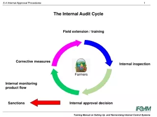
Outline • R & D permission obtained • Phase 1 • Assess current practice of CVC care in relation to prevention of VAE in all locations throughout the hospital • Presentation of results • Phase 2 • Assess need for standard setting • Implement agreed standard • Phase 3 • Audit at 1 & 6 months post intervention
Data collection proforma Audit ID number: Location: Bed number: CVC Site R L IntJug Subclavian Femoral Lumens in total 1 2 3 4 5 Lumens in use 1 2 3 4 5 Reason for CVC Speciality/Grade of Dr inserting line Insertion date Sutures Fixed connector sutured Y N Adjustable connector present Y N & sutured Y N Comments Dressing Covering insertion site Y N Clean Y N What position should the patient be in when removing the CVC? (ask nurse looking after patient) Bung Bionector Tap position Clip open Clip Closed X Leave blank if no clip If single bionector attached to lumen then write BIONECTOR across diagram
Data collection • Wed 28th Oct 2009 • all wards in hospital • ICU, HDU, CICU, CCU, medical & surgical wards, PACUs. • all patients with CVC in situ included in the evaluation • data collection proforma completed for each CVC
Results: common errors 3 way Tap CVC lumen Patient Patient IVI IVI Patient Patient
Intervention • presentation locally • raised awareness • ensure CVC chosen is appropriate • use of three-way taps? • hospital standard set • re-education • Poster
Prevention of Venous Air Embolism (VAE): Central Venous Catheter (CVC) Care 1. Service evaluation Oct 09: 2. Intervention: Points to remember CVC insertion site: CVC sutured to the skin at all times Insertion site covered by an occlusive dressing % of CVCs with errors potentially leading to VAE Prevent air from entering CVC: Prime all syringes & IV giving sets Use needle-free access devices if possible Ensure bungs are not cross-threaded Ensure correct 3-way-tap alignment: ✓ ✓ 64% of CVCs had an error 64% of CVCs at risk of VAE Removal: Follow trust guidelines but remember to: Lie the patient head down Apply a sterile occlusive dressing 3. Re-audit planned summer 2010 ✗ ✗ ✗
What next? • repeat education / updated posters
Prevention of Venous Air Embolism (VAE): Central Venous Catheter (CVC) Care 1. Current practice: 2. Intervention: Points to remember CVC insertion site: CVC sutured to the skin at all times Insertion site covered by an occlusive dressing % of CVCs with errors potentially leading to VAE Prevent air from entering CVC: Prime all syringes & IV giving sets Use needle-free access devices if possible Ensure bungs are not cross-threaded Ensure correct 3-way-tap alignment: ✓ ✓ Removal: Follow trust guidelines but remember to: Lie the patient head down Apply a sterile occlusive dressing Oct 09: 64% of CVCs at risk of VAE May 10: 35% of CVCs at risk of VAE 3. Re-audit planned Nov 2010 ✗ ✗ ✗
What next? • repeat education / updated posters • needle-less valves? • re-audit 6 months
Summary • raised awareness relating to VAE • prevention • management • our hospital’s approach • consider… • need for CVC? • lumens required? • needle-free valves?