Download
1 / 105
1.11k likes | 1.5k Vues
Nerve Cells. Nervous System. Three Parts of the Nervous System Central Nervous System ( CNS ): brain and spinal cord. Peripheral Nervous System ( PNS ): nerves of the body

E N D
Nervous System • Three Parts of the Nervous System • Central Nervous System (CNS): brain and spinal cord. • Peripheral Nervous System (PNS): nerves of the body • Autonomic Nervous System (ANS): has parts of the CNS and PNS. Controls autonomic function (blood pressure, digestion, etc). • Sympathetic division • Parasympathetic division
Basic Divisions of the Nervous System Figure 12.2
Basic Divisions of the Nervous System • Central nervous system (CNS) • Brain and spinal cord • Integrating and command center
Basic Divisions of the Nervous System • Peripheral nervous system (PNS) • Outside the CNS • Consists of nerves from brain and spinal cord • Cranial nerves • Spinal nerves • Peripheral nerves link all regions of the body to the CNS
Neurons • The nervous system is made up of more cells than any other system. • For instance, the brain has about 100 billion cells. • There are also a number of different cell types, the most important is the neuron.
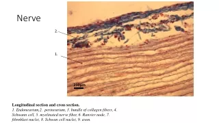
Structure of a Nerve The axon of each neuron is surrounded by a sheath called the endoneurium. Some axons have an additional sheath called myelin. A bundle of neurons travel together in a fascicle, and are surrounded by perineurium. A bundle of fascicles is surrounded by epineurium Figure 12.16a
NEURON • All neurons do three things: • Receive a signal. Can be any type of stimulus (touch, vibration, light, sound, signal from another neuron, etc). • Transmit a signal to another location. E.g. finger touching something signal to spinal cord or brain. • Stimulate another cell • Another neuron transmit signal • Muscle contraction • Gland secretion • Blood vessel constriction
The Neuron • Other special characteristics • Longevity – can live and function for a lifetime • Do not divide – fetal neurons lose their ability to undergo mitosis because their centrioles no longer function and cannot pull the chromosomes apart; neural stem cells are an exception • High metabolic rate – require abundant oxygen and glucose
Classification of Neurons • Structure (Appearance) • Unipolar • Bipolar • Multipolar • Function • Sensory • Motor • Interneurons
Structural Classification of Neurons Multipolar neurons • Most neurons are this type, having many dendrites and one axon. Bipolar neurons • Have two processes that extend from opposite sides of the cell body. Some sensory neurons are bipolar. Unipolar neurons Have one short process emerging from the cell body, which then branches into a “T”. Sensory neurons in the PNS are unipolar.
Neurons are grouped functionally according to the direction the nerve impulse travels relative to the CNS. Sensoroy Neurons (afferent neurons) transmit impulses toward the CNS. They originate in the PNS and terminate in the CNS. Motor Neurons (efferent neurons) transmit impulses from the CNS to effector organs (muscles and glands). They originate in the CNS and terminate in the PNS. Interneurons (association neurons) connect sensory neurons to motor neurons within the spinal cord and brain. They originate and terminate in the CNS, and form complex neuronal pathways. They make up 99.98% of the neurons in the body, reflecting the vast amount of information processed in the CNS. Functional Classification of Neurons
Neurons Classified by Function: Sensory vs. Motor Neurons Sensory neurons enter the spinal cord. Motor neurons leave the spinal cord. Interneurons connect the sensory and motor neurons. Figure 12.11
There are hundreds of different types of neurons, each one is specialized for a particular task • (e.g. sensory neurons receive and transmit sensory information, and there are several different types of them, with receptors for light, smell, pain, light touch, vibration, position in space). • Motor neurons transmit signals for muscle contraction (organ contraction, blood vessel constriction), gland secretion. • They all share certain characteristics. • Longevity (can last a lifetime) • High metabolic rate • Cannot divide to reproduce (they lose their centrioles during fetal development) • Cannot survive without oxygen
Neuron Anatomy Neuron Anatomy Soma (cell body) Axon terminals (stimulate another cell) Axon (transmits signals) Axon hillock (trigger zone) Dendrites (receive signal)
NEUROLEMMA is the name of the plasma membrane (outermost covering) of a neuron. • DENDRITES function to receive the signal and carry the nerve conduction toward the cell body. • SOMA (cell body) is where the nucleus, ribosomes, and most organelles are located • AXON HILLOCK is the area on the soma where the action potential (electrical charges) of the neuron builds up before it transmits the signal down the axon. • AXON function is to transmit signals. Some cells have many axons, some have one, some are short, and some are long. • AXON TERMINALS (also called boutons or synaptic knobs) contain a neurotransmitter which, when released, stimulates another cell. • A SYNAPSE is where one neuron touches another neuron. Neurons may have a couple of synapses, or hundreds. • AXOPLASMIC TRANSPORT: Movement of nutrients, wastes, and organelles between the cell body and axon terminals
Nerve Impulses (action potentials) • A nerve impulse (called an action potential) is typically generated at the axon hillock, and is conducted along the axon to the axon terminals, where it causes the release of neurotransmitters into the extracellular space. • These neurotransmitters excite or inhibit the dendrites of the adjacent neuron (or the target organ). • If they excite the dendrites of an adjacent neuron, the action potential is carried to the cell body of the second neuron, and then it continues down the axon to excite a third neuron, and so on, until the target organ is reached.
Structure of a Typical Large Neuron Figure 12.4
A synapse is the site at which two neurons communicate, or a neuron and its target organ. The neuron that conducts the signals towards the synapse is called the presynaptic neuron. The neuron that transmits the signals away from the synapse is called the postsynaptic neuron. Most neurons function as pre-synaptic and post-synaptic neurons. The neurons don’t physically touch each other at the synapse. The space between them is called the synaptic cleft. Synapse
Two Neurons Communicating at a Synapse Know this order: The action potential travels from the Axon of presynaptic neuron SYNAPTIC CLEFT dendrite of post synaptic neuron Figure 12.6
Types of Synapses • Axosomatic (axon touches soma) • Axodendritic (axon touches dendrite) • Axoaxonic (axon touches another axon)
Axosomatic Synapse Figure 12.7
How does the signal go through the space? By a chemical transmission. • The axon terminals have vesicles filled with a neurotransmitter that transmits the signal across the synapse. • Each type of neuron uses a particular type of neurotransmitters, so there are many types of neurotransmitters. • Some neurotransmitters excite the adjacent neuron, and some are inhibitory.
Structure of a Synapses Chemical substances released from the presynaptic terminal: • May inhibit or stimulate an action in the postsynaptic cell • May be broken down by enzymes or taken back up into the vesicles and recycled. Figure 12.8a, b
Fun Fact • Children under 3 years of age have twice as many synapses as adults. That is why they learn languages better. • A child must learn a second language before the age of ten, or they probably will not have the proper native accent of that language.
The axon terminal of the nerve cell rests in indentations in the cell membrane of the muscle fiber. • The enlarged knob of the axon is called the presynaptic terminal • The space between the presynaptic terminal and the muscle fiber membrane is the synaptic cleft • The muscle fiber membrane is the postsynaptic membrane. • If this neuron innervates skeletal muscle, the vesicles of its axon terminal will contain the neurotransmitter acetylcholine.
An action potential causes the release of Ach (acetylcholine; the neurotransmitter at the neuromuscular junction) into the synaptic cleft. • Ach binds to receptor sites on the muscle fiber (muscle cell) membrane and starts an electrical impulse called an action potential, which travels along the length of the muscle fiber and causes it to contract. • The Ach that was released is rapidly broken down by an enzyme, Ach-ase (acetylcholinesterase). This ensures that the action potential will result in only one contraction of each muscle fiber. • A neuron might be temporarily unable to transmit an impulse to another cell ifits supply of neurotransmitters is exhausted.
AchE Blockers • Neostigmine • Physostigmine • Myasthenia Gravis-ptosis
Myasthenia gravis Myasthenia gravis is an autoimmune disorder in which antibodies attack and destroy some acetylcholine receptors. Acetylcholine is therefore less likely to stimulate muscle contraction, resulting in muscle weakness and fatigue. Symptoms usually begin in the eyelid and facial muscles, and manifests as drooping muscles on half or both sides of the face, drooping eyelids, and slurred speech. Neostigmine is an anti-cholinesterase drug which reduces the symptoms by inhibiting Ach-ase activity, preventing the breakdown of Ach. Consequently, Ach levels in the synapse remain elevated, so Ach is available to bind to those few functional Ach receptors that are left.
AchE irreversible inhibitor • Sarin gas • Chemical warfare • Spastic paralysis • Ventilator until new AchE can be made
Acetylcholine-esterase Blocker Acetylcholine Antagonists Some INSECTICIDES inhibit acetylcholinesterase, so Ach accumulates in the synaptic cleft and acts as a constant stimulus to the muscle fiber. The insects die because their respiratory muscles contract and cannot relax. Other poisons, such as CURARE, the poison used by South American Indians in poison arrows, bind to the Ach receptors on the muscle cell membrane and prevent Ach from working. That prevents muscle contraction, resulting in flaccid paralysis.
Inhibitory Neuron Blockers • Tetanus exotoxin • Blocks release of inhibitory neurotransmitters • Muscles can’t relax • Spastic paralysis • Opposing flexor and extensor muscles contract
Cell Membranes • What two conditions must be met for diffusion of a substance across a semipermeable membrane? • Is the membrane permeable to it? • Does it have a concentration gradient? • If the answer is yes to both questions, then the substance will diffuse (Which way? Down it’s gradient)
Each cell in our body is surrounded by a cell membrane composed of a phospholipid BI-LAYER. • That means that our cell membranes have two layers: an outer layer, and an inner layer. • The inside layer of each cell membrane in the body, (including each neuron) has a charge (usually negative), and the outside layer of each cell membrane has a charge (usually positive).
The reason for the charge difference is that there are many proteins inside of cells, and proteins are made of amino acids, most of which have a negative charge. Because proteins are negatively charged, the inside layer of the cell membrane has a negative charge. • Outside of the cell, there are many electrolytes, especially sodium (Na+), which have a positive charge. They stay outside of the cell because they cannot get in unless sodium channels in the cell membrane are open, which they usually are not. That’s why the outside of the cell membrane usually has a positive charge.
What is a sodium channel? Proteins embedded in the cell membranes form channels which only allow certain ions to cross the cell membrane. • A sodium ion can only get into the cell by way of a sodium channel. A potassium ion can only get in by way of a potassium channel, etc. • Charged ions, such as potassium (K+), sodium (Na+), calcium (Ca++), and chloride (Cl-) are called electrolytes. When they move from one side of the cell membrane to the other (when their channels are open), they carry their electrical charge with them. • This changes the overall charge of the inside and outside of the cell membrane.
Not just separation of solutes, but charges, too! + + + + • Inside of the cell is negative due to : • Abundance of proteins, which have a negative charge • The cell membrane is very permeable (“leaky”) to K+, so it can LEAVE the cell whenever it wants. That leaves the inside of the cell more negative. _ _ _ _ _ _ _ _ _ _ _ _ + + + + + + + + + + + +
Sidedness “Sidedness” of the membrane • Sidedness means that the electrical charges on one side of the membrane (positive or negative) are different than on the other side. Why does sidedness exist? • The cell membrane has different permeabilities to each ion; for instance the cell is more permeable to K+ than any other ion. • Pumps exist which force particular ions into or out of the cell • Channels made out of protein selectively allow particular ions into or out of the cell. These channels may be open or closed at any given time.
Every cell has a positive charge on the outside of the membrane and a negative charge on the inside of the cell membrane, when the cell is at rest (not being stimulated). • K+ constantly leaks out of the cell because it wants to diffuse down its concentration gradient. That means it wants to go from its area of high concentration (the inside of the cell) to an area where it is in low concentration (the outside of the cell). • Na+ wants to get into the cell, too, but it’s channel is closed. • Because of this separation of chemicals and electrical charges, every cell has a Resting Membrane “Potential”. • There is a difference in electrical charge across the membrane (a potential difference) • More negative inside; more positive outside • Resting membrane potential is minus70 to minus 90mV
As K+ leaves the cell, it takes a positive charge outside with it, so the inside is more negative. • However, as the inside of the cell is becoming more negative, the outside of the cell is becoming more positive, and the positive charges will want to flow back inside of the cell since they are attracted to the negative charges. • This is what keeps K+ from just leaving the cell until it is in equal numbers on both sides of the cell. Before it can reach such an equilibrium, it gets pulled back into the cell because its positive charges are drawn into the inside of the cell, where the charge has become strongly negative (because proteins are on the inside of the cell and they have a negative charge). • Other positively charged ions, like Na+, want to go into the cell also, but they are blocked by protein gates that only K+ can get through.
ACTION POTENTIAL • The resting membrane potential • Occurs because the cell membrane is more permeable to potassium ions than sodium ions • Occurs because there are negatively charged proteins and ions inside the cell • The action potential occurs if the membrane potential reaches a certain threshold.
Na+ Cl- Cl- Na+ + + Na+ Cl- + + Cl- Na+ - - - - Na+ Cl- Na+ Cl- Na+ Cl- -70 mV What if….. • What if a membrane suddenly became MORE PERMEABLE to Na+????? • Even for just a moment in time….. • What would Na+ do? (Ask yourself the 3 questions) Which way is the electrochemical gradient for Na+? Electrical: inward Chemical: inward Answer: Most definitely INWARD Sodium WANTS IN! Can it get in? What would happen to the membrane potential of the cell when this event occurs?
Know the following: • Direction of impulse • Ions involved, charges • States: resting potential, depolarization, repolarization Na+ Na+ Na+ Resting potential Depolarization Repolarization Na+ enters and K+ leaves, so outside of membrane becomes negative charge. This is depolarization When stimulus is over, Na+ leaves and K+ lenters the cell, so outside of membrane returns to positive charge. This is repolarization Outside of membrane is positive charge at resting potential. Direction of impulse
What would happen to the membrane potential of the cell when you open up a sodium channel? • If we instantly increase sodium permeability, sodium will enter the cell, changing the charge of the inside of the cell so that it goes from negative to positive. The outside layer of the cell membrane would then go from positive to negative (the charges flip). This is called DEPOLARIZATION. • However, when this occurs, Na+ will be in higher concentration on the inside of the cell, so it wants to diffuse back out of the cell. • Once it leaves the cell again, the membrane potential of the inside of the cell membrane will return to a negative charge. This is called REPOLARIZATION.
Direction of Depol Resting Cell + + + + + + + + + + + + + + + + + + + + + + + + + + + + + + + + - - - - - - - - - - - - - - - - - - - - - - - - - - - - - - - - - - - - - - - - - - - - - - - - - - - - - - - - - - - - - - - - - - - - - - - - - - - - - - - - - - - - - - + + + + + + + + + + + + + + + + + + + + + + + + + + + + + + + +
Direction of Depol + + + + + + + + + + + + + + + + + + + + + + + + + + + + + + + + - - - - - - - - - - - - - - - - - - - - - - - - - - - - - - - - - - - - - - - - - - - - - - - - - - - - - - - - - - - - - - - - - - - - - - - - - - - - - - - - - - - - - - + + + + + + + + + + + + + + + + + + + + + + + + + + + + + + + + Sodium channels open
Direction of Depol + + + + + + + + + + + + + + + + + + + + + + + + + + + + + + + + - - - - - - - - - - - - - - - - - - - - - - - - - - - - - - - - - - - - - - - - - - - - - - - - - - - - - - - - - - - - - - - - - - - - - - - - - - - - - - - - - - - - - - + + + + + + + + + + + + + + + + + + + + + + + + + + + + + + + +