Download

1 / 61
1.75k likes | 4.43k Vues
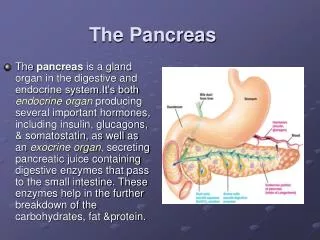
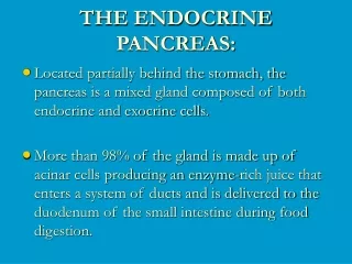
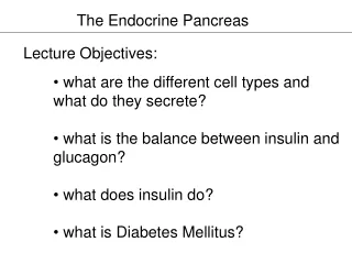
The Endocrine Pancreas. Regulation of Carbohydrate Metabolism. Pancreatic Anatomy. Gland with both exocrine and endocrine functions 15-25 cm long 60-100 g Location: retro-peritoneum, 2 nd lumbar vertebral level Extends in an oblique, transverse position

E N D
The Endocrine Pancreas Regulation of Carbohydrate Metabolism
Pancreatic Anatomy Gland with both exocrine and endocrine functions 15-25 cm long 60-100 g Location: retro-peritoneum, 2nd lumbar vertebral level Extends in an oblique, transverse position Parts of pancreas: head, neck, body and tail
Head of Pancreas Includes uncinate process Flattened structure, 2 – 3 cm thick Attached to the 2nd and 3rd portions of duodenum on the right Emerges into neck on the left Border b/w head and neck is determined by GDA insertion SPDA and IPDA anastamose between the duodenum and the right lateral border
Neck of Pancreas 2.5 cm in length Straddles SMV and PV Antero-superior surface supports the pylorus Superior mesenteric vessels emerge from the inferior border Posteriorly, SMV and splenic vein confluence to form portal vein Posteriorly, mostly no branches to pancreas
Body of Pancreas Elongated, long structure Anterior surface, separated from stomach by lesser sac Posterior surface, related to aorta, lt. adrenal gland, lt. renal vessels and upper 1/3rd of lt. kidney Splenic vein runs embedded in the post. Surface Inferior surface is covered by transverse mesocolon
Tail of Pancreas Narrow, short segment Lies at the level of the 12th thoracic vertebra Ends within the splenic hilum Lies in the splenophrenic ligament Anteriorly, related to splenic flexure of colon May be injured during splenectomy (fistula)
Pancreatic Duct Main duct (Wirsung) runs the entire length of pancreas Joins CBD at the ampulla of Vater 2 – 4 mm in diameter, 20 secondary branches Ductal pressure is 15 – 30 mm Hg (vs. 7 – 17 in CBD) thus preventing damage to panc. duct Lesser duct (Santorini) drains superior portion of head and empties separately into 2nd portion of duodenum
Arterial Supply of Pancreas Variety of major arterial sources (celiac, SMA and splenic) Celiac Common Hepatic Artery Gastroduodenal Artery Superior pancreaticoduodenal artery which divides into anterior and posterior branches SMA Inferior pancreaticoduodenal artery which divides into anterior and posterior branches
Arterial Supply of Pancreas • Anterior collateral arcade between anterosuperior and anteroinferior PDA • Posterior collateral arcade between posterosuperior and posteroinferior PDA • Body and tail supplied by splenic artery by about 10 branches • Three biggest branches are • Dorsal pancreatic artery • Pancreatica Magna (midportion of body) • Caudal pancreatic artery (tail)
Venous Drainage of Pancreas • Follows arterial supply • Anterior and posterior arcades drain head and the body • Splenic vein drains the body and tail • Major drainage areas are • Suprapancreatic PV • Retropancreatic PV • Splenic vein • Infrapancreatic SMV • Ultimately, into portal vein
Lymphatic Drainage • Rich periacinar network that drain into 5 nodal groups • Superior nodes • Anterior nodes • Inferior nodes • Posterior PD nodes • Splenic nodes
Innervation of Pancreas Sympathetic fibers from the splanchnic nerves Parasympathetic fibers from the vagus Both give rise to intrapancreatic periacinar plexuses Parasympathetic fibers stimulate both exocrine and endocrine secretion Sympathetic fibers have a predominantly inhibitory effect
Innervation of Pancreas Peptidergic neurons that secrete amines and peptides (somatostatin, vasoactive intestinal peptide, calcitonin gene-related peptide, and galanin Rich afferent sensory fiber network Ganglionectomy or celiac ganglion blockade interrupt these somatic fibers (pancreatic pain)
Pancreatic Hormones, Insulin and Glucagon, Regulate Metabolism
Production of Pancreatic Hormones by Three Cell Types • Alpha cells produce glucagon. • Beta cells produce insulin. • Delta cellsproduce somatostatin.
Islet of Langerhans Cross-section • Three cell types are present, A (glucagon secretion), B (Insulin secretion) and D (Somatostatin secretion) • A and D cells are located around the perimeter while B cells are located in the interior • Venous return containing insulin flows by the A cells on its way out of the islets
Pancreatic Hormones, Insulin and Glucagon, Regulate Metabolism Figure 22-8: Metabolism is controlled by insulin and glucagon
Structure of Insulin • Insulin is a polypeptide hormone, composed of two chains (A and B) • BOTH chains are derived from proinsulin, a prohormone. • The two chains are joined by disulfide bonds.
Roles of Insulin • Acts on tissues (especially liver, skeletal muscle, adipose) to increase uptake of glucose and amino acids. - without insulin, most tissues do not take in glucose and amino acids well (except brain). • Increases glycogen production (glucose storage) in the liver and muscle. • Stimulates lipid synthesis from free fatty acids and triglycerides in adipose tissue. • Also stimulates potassium uptake by cells (role in potassium homeostasis).
phosphorylation of insulin responsive substrates (IRS) RAS RAF-1 MAP-K MAP-KK Final actions The Insulin Receptor • The insulin receptor is composed of two subunits, and has intrinsic tyrosine kinase activity. • Activation of the receptor results in a cascade of phosphorylation events:
Specific Targets of Insulin Action: Carbohydrates • Increased activity of glucose transporters. Moves glucose into cells. • Activation of glycogen synthetase. Converts glucose to glycogen. • Inhibition of phosphoenolpyruvate carboxykinase. Inhibits gluconeogenesis.
lipoprotein lipase Specific Targets of Insulin Action: Lipids • Activation of acetyl CoA carboxylase. Stimulates production of free fatty acids from acetyl CoA. • Activation of lipoprotein lipase (increases breakdown of triacylglycerol in the circulation). Fatty acids are then taken up by adipocytes, and triacylglycerol is made and stored in the cell.
Regulation of Insulin Release • Major stimulus: increased blood glucose levels - after a meal, blood glucose increases - in response to increased glucose, insulin is released - insulin causes uptake of glucose into tissues, so blood glucose levels decrease. - insulin levels decline as blood glucose declines
Insulin Action on Cells: Dominates in Fed State Metabolism • glucose uptake in most cells (not active muscle) • glucose use and storage • protein synthesis • fat synthesis
Other Factors Regulating Insulin Release • Amino acids stimulate insulin release (increased uptake into cells, increased protein synthesis). • Keto acids stimulate insulin release (increased glucose uptake to prevent lipid and protein utilization). • Insulin release is inhibited by stress-induced increase in adrenal epinephrine - epinephrine binds to alpha adrenergic receptors on beta cells - maintains blood glucose levels • Glucagon stimulates insulin secretion (glucagon has opposite actions).
Structure and Actions of Glucagon • Peptide hormone, 29 amino acids • Acts on the liver to cause breakdown of glycogen (glycogenolysis), releasing glucose into the bloodstream. • Inhibits glycolysis • Increases production of glucose from amino acids (gluconeogenesis). • Also increases lipolysis, to free fatty acids for metabolism. • Result: maintenance of blood glucose levels during fasting.
Mechanism of Action of Glucagon • Main target tissues: liver, muscle, and adipose tissue • Binds to a Gs-coupled receptor, resulting in increased cyclic AMP and increased PKA activity. • Also activates IP3 pathway (increasing Ca++)
Glucagon Action on Cells: Dominates in Fasting State Metabolism • Glucagon prevents hypoglycemia by cell production of glucose • Liver is primary target to maintain blood glucose levels
Glucagon Action on Cells: Dominates in Fasting State Metabolism
Targets of Glucagon Action • Activates a phosphorylase, which cleaves off a glucose 1-phosphate molecule off of glycogen. • Inactivates glycogen synthase by phosphorylation (less glycogen synthesis). • Increases phosphoenolpyruvate carboxykinase, stimulating gluconeogenesis • Activates lipases, breaking down triglycerides. • Inhibits acetyl CoA carboxylase, decreasing free fatty acid formation from acetyl CoA • Result: more production of glucose and substrates for metabolism
Regulation of Glucagon Release • Increased blood glucose levels inhibit glucagon release. • Amino acids stimulate glucagon release (high protein, low carbohydrate meal). • Stress: epinephrine acts on beta-adrenergic receptors on alpha cells, increasing glucagon release (increases availability of glucose for energy). • Insulin inhibits glucagon secretion.
Other Factors Regulating Glucose Homeostasis • Glucocorticoids (cortisol): stimulate gluconeogenesis and lipolysis, and increase breakdown of proteins. • Epinephrine/norepinephrine: stimulates glycogenolysis and lipolysis. • Growth hormone: stimulates glycogenolysis and lipolysis. • Note that these factors would complement the effects of glucagon, increasing blood glucose levels.
Hormonal Regulation of Nutrients • Right after a meal (resting): • - blood glucose elevated • - glucagon, cortisol, GH, epinephrine low • - insulin increases (due to increased glucose) • - Cells uptake glucose, amino acids. • - Glucose converted to glycogen, amino acids into protein, lipids stored as triacylglycerol. • - Blood glucose maintained at moderate levels.
Hormonal Regulation of Nutrients • Afew hours after a meal (active): • - blood glucose levels decrease • - insulin secretion decreases • - increased secretion of glucagon, cortisol, GH, epinephrine • - glucose is released from glycogen stores (glycogenolysis) • - increased lipolysis (beta oxidation) • - glucose production from amino acids increases (oxidative deamination; gluconeogenesis) • - decreased uptake of glucose by tissues • - blood glucose levels maintained
Turnover Rate • Rate at which a molecule is broken down and resynthesized. • Average daily turnover for carbohydrates is 250 g/day. • Some glucose is reused to form glycogen. • Only need about 150 g/day. • Average daily turnover for protein is 150 g/day. • Some protein may be reused for protein synthesis. • Only need 35 g/day. • 9 essential amino acids. • Average daily turnover for fats is 100 g/day. • Little is actually required in the diet. • Fat can be produced from excess carbohydrates. • Essential fatty acids: • Linoleic and linolenic acids.
Regulation of Energy Metabolism • Energy reserves: • Molecules that can be oxidized for energy are derived from storage molecules (glycogen, protein, and fat). • Circulating substrates: • Molecules absorbed through small intestine and carried to the cell for use in cell respiration. Insert fig. 19.2
Pancreatic Islets (Islets of Langerhans) • Alpha cells secrete glucagon. • Stimulus is decrease in blood [glucose]. • Stimulates glycogenolysis and lipolysis. • Stimulates conversion of fatty acids to ketones. • Beta cells secrete insulin. • Stimulus is increase in blood [glucose]. • Promotes entry of glucose into cells. • Converts glucose to glycogen and fat. • Aids entry of amino acids into cells.
Energy Regulation of Pancreas • Islets of Langerhans contain 3 distinct cell types: • a cells: • Secreteglucagon. • b cells: • Secreteinsulin. • D cells: • Secrete somatostatin.
Regulation of Insulin and Glucagon • Mainly regulated by blood [glucose]. • Lesser effect: blood [amino acid]. • Regulated by negative feedback. • Glucose enters the brain by facilitated diffusion. • Normal fasting [glucose] is 65–105 mg/dl.
Regulation of Insulin and Glucagon (continued) • When blood [glucose] increases: • Glucose binds to GLUT2 receptor protein in b cells, stimulating the production and release of insulin. • Insulin: • Stimulates skeletal muscle cells and adipocytes to incorporate GLUT4 (glucose facilitated diffusion carrier) into plasma membranes. • Promotes anabolism.
Oral Glucose Tolerance Test • Measurement of the ability of b cells to secrete insulin. • Ability of insulin to lower blood glucose. • Normal person’s rise in blood [glucose] after drinking solution is reversed to normal in 2 hrs. Insert fig. 19.8
Regulation of Insulin and Glucagon • Parasympathetic nervous system: • Stimulates insulin secretion. • Sympathetic nervous system: • Stimulates glucagon secretion. • GIP: • Stimulates insulin secretion. • GLP-1: • Stimulates insulin secretion. • CCK: • Stimulates insulin secretion.
Bodycellstake up moreglucose Insulin Beta cellsof pancreas stimulatedto release insulin intothe blood • Glucose homeostasis – Putting it all together Liver takesup glucoseand stores it asglycogen Blood glucose leveldeclines to a set point;stimulus for insulinrelease diminishes High bloodglucose level STIMULUS:Rising blood glucoselevel (e.g., after eatinga carbohydrate-richmeal) Homeostasis: Normal blood glucose level(about 90 mg/100 mL) STIMULUS:Declining bloodglucose level(e.g., afterskipping a meal) Blood glucose levelrises to set point;stimulus for glucagonrelease diminishes Alphacells ofpancreas stimulatedto release glucagoninto the blood Liverbreaks downglycogen and releases glucoseto the blood Glucagon Figure 26.8
Hormonal Regulation of Metabolism • Absorptive state: • Absorption of energy. • 4 hour period after eating. • Increase in insulin secretion. • Postabsorptive state: • Fasting state. • At least 4 hours after the meal. • Increase in glucagon secretion.