Download
1 / 40
400 likes | 470 Vues
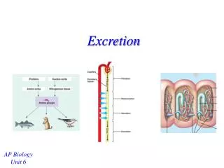
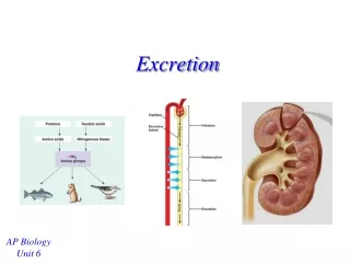
Mammal Excretion and Reproduction. Ch 49 &. Urinary system. Kidney Renal cortex Renal medulla Inner region Outer region Ureters Urinary bladder Urethra. Nephron. Functional unit of kidney: forms urine Renal corpuscle forms filtrate Tubule performs secretion and reabsorption

E N D
Urinary system • Kidney • Renal cortex • Renal medulla • Inner region • Outer region • Ureters • Urinary bladder • Urethra
Nephron Functional unit of kidney: forms urine Renal corpuscle forms filtrate Tubule performs secretion and reabsorption Tubule empties into collecting duct Excretion Fig 17.2 17-10
Renal corpuscle • Glomerulus • Cluster of interconnected, fenestrated capillaries • Supplied by afferent arteriole • Drained by efferent arteriole • Bowman’s capsule • Podocytes form filtration slits (physical barriers)
Fig 17.5 17-13
Filtration • 20% of plasma leaves glomerulus and filters into Bowman’s space • Proteins and blood cells remain in plasma • Glomerular filtrate • Glomerular filtration Rate (GFR): rate of filtrate production
Reabsorption in PCT • Water and solutes • Na+, K+, Cl-, HCO3-, glucose, amino acids 17-13
Reabsorption in Loop of Henle • Water reabsorption • Loop sets up a concentration gradient between tissues and tubule • Active transport (AT) of salt out of ascending loop • Water moves by osmosis in descending loop
Ascending Limb LH Impermeable to H20; permeable to salt; thick part ATs salt out of filtrate AT of salt causes filtrate to become dilute (100 m O sm) by end of LH Fig 17.17 17-38
Descending Limb LH Permeable to H20 Impermeable to salt Deep regions of medulla are 1400 m O sm, H20 diffuses out of filtrate until it equilibrates with interstitial fluid H20 reabsorbed by capillaries 17-37
Desert gerbil that concentrates urine to a high degree. • Long loops of Henle increase the concentration gradient in the surrounding tissues. • The desert gerbil does not drink!
Distal Convoluted Tubule • Secretion of K+, H+ • Absorption of Na+ • Aldosterone (hormone) • Stimulates active transport of 3 molecules of Na+ out of tubule (reabsorption) for every 2 molecules of K + brought into tubule (secretion) • Water from tubule follows Na+ by osmosis into blood (reabsorption of water)
Collecting Duct • Uses gradient set up by loop of Henle determine final urine volume • Antidiuretic hormone (ADH) • Causes an increase the number of aquaporins in the collecting duct membranes • Higher levels of ADH increase the number of aquaporins allowing water to be reabsorbed: urine volume decreases
Transport maximum (Tm) • Reabsorption will return solutes to the blood • Binding sites for transport can become saturated at high levels • Solutes not reabsorped are lost in the urine • Vitamin C • Glucose
Mammalian reproductive structure and function Male Genitalia: penis and scrotum (testes) Testes 2°C lower than core body temperature Seminiferous tubules and Leydig cells (secrete testosterone) Sertoli cells provide nutrients and protection to developing sperm 20
Fig 27.1
Fig 27.7
Fig 27.5 There is about one mile of seminiferous tubule in men This allows men to produce about 500,000,000 sperm/day
Epididymis: sperm complete differentiation Vas deferens ejaculatory duct urethra Semen Sperm: 5% of volume Fluid from seminal vesicles (fructose), bulbourethral glands (alkaline mucus), and prostate gland (protective fluid) 24
Hypothalamus Gonadotropin-releasing hormone (GnRH) Anterior pituitary luteinizing hormone (LH) and follicle-stimulating hormone (FSH) Leydig cells produce testosterone Sertoli cells and germ cells to stimulate spermatogenesis Controlled by negative feedback Increased GnRH at puberty initiates release of LH and FSH Testosterone secretion 26
Female Vagina cervix uterus Endometrium: mucous membrane Myometrium: muscular layer 28
Ovaries: egg development One egg released into abdominal cavity Drawn into oviduct or fallopian tube by fimbriae Oviduct (fallopian tube) Fertilization Blastocyst: ball of 32-150 cells that enters uterus 30
Oogenesis Most female mammals produce all 1° oocytes before birth 1 million at birth 200,000 in each ovary at puberty Ovarian cycle ~28 days in humans Several oocytes begin maturation only 1 is ovulated each cycle Menopause- oocytes become depleted and ovulation stops 32
Ovarian cycle Week 1: several 1° oocytes begin to develop, each within a follicle Week 2: 1 follicle continues developing 1° oocyte 2° oocyte 2° oocyte surrounded by cumulus mass-- secretes estradiol meiosis 33
Ovarian cycle Follicle matures with2o oocyte Ovulation: follicle ruptures releasing egg, zona pellucida, and cumulus mass Corpus luteum: secretes progesterone No pregnancy: corpus luteum degenerates to corpus albicans 34
Follicular phase First half Growth and differentiation of follicle Low LH levels stimulate follicular cells to make estradiol Estradiol levels slowly increase Mature follicle: estradiol production jumps Positive feedback: LH spike Ovulation Spontaneous vs. induced ovulation Luteal phase Corpus luteum secretes progesterone Inhibits FSH and LH secretion prepares uterus Fertilization: cells surrounding embryo produce chorionic gonadotropin-- maintains corpus luteum No fertilization: corpus luteum degenerates in 2 weeks and cycle begins again 36
Menstruation starts corpus luteum degenerates Other mammals have estrous cycles Proliferative phase Secretory phase Implantation: pregnancy begins No implantation, lining shed Uterine (menstrual) cycle 37
Pregnancy or gestation Developing embryo grows within the uterus of the mother Physiologically, begins when the embryo is established in the uterine lining Length varies widely and is roughly related to adult size Elephant: 22 months Shrew: 45 - 60 days 39
African elephant Elephant shrew 40