Download
1 / 42
420 likes | 568 Vues
Prepared by: Bishly. Case Study. Demographic Data. CASE NO: 14xxx NAME: Baby H AGE: 3 yrs old SEX: Male DIAGNOSIS: Intussusception. Physical Assessment. Vital Signs: BP- 90/60 HR- 126 bpm RR- 26/ min T- 38.5 C SPO2- 98%. General Assessment.

E N D
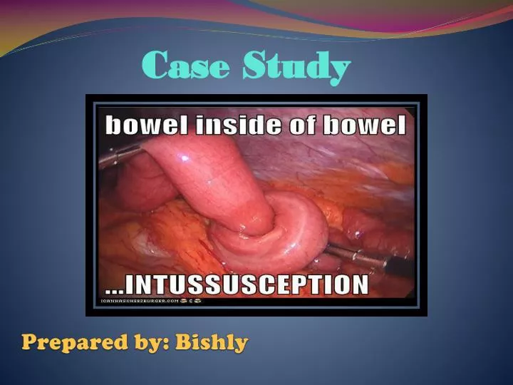
Prepared by: Bishly Case Study
Demographic Data • CASE NO: 14xxx • NAME: Baby H • AGE: 3 yrs old • SEX: Male • DIAGNOSIS: Intussusception
Vital Signs: • BP- 90/60 • HR- 126 bpm • RR- 26/ min • T- 38.5 C • SPO2- 98%
General Assessment • Patient is conscious, coherent and oriented. • Cries intermittently and recovers from pain Skin • No lesion, moist and good turgor • Pale in appearance
Head and Neck • No palpable lesion on the head and neck.
Thorax • Trachea in central • Chest movement are equal • Air entry is equal in all areas, no added sound
Abdomen • No distention, no dilated veins • Guarding present, no rigidity in the right hypochondrium • Tenderness on the right hypochondrium • No free fluid on percussion • Bowel sound is present
Genitalia • Circumcised • Testes are bilateral descended and normal in size Neurological • No neurologic defect seen
Past Medical History • The patient has no major medical problems • No history of surgery
Present Medical History • Patient’s chief complaints include colicky abdominal pain associated with fever for 2 days • Patient was seen by Dr. Jacob and was advised for ultrasound • On examination: Febrile (Temp- 38 C) RR-20/ min SPO2- 97 % WT- 13 kg PR- 112 bpm Child cries intermittently and recovers from the pain Ultrasound done (shows “doughnut- like image”-target sign)
Topic presentation INTUSSUSCEPTION
Introduction • Intussusception is a term derived from the Latin intus(within) and suscipere(to receive). One segment of the bowel (intussusceptum) invaginates into another (intussuscepien) just distal to it, much like the piece of a telescope. When this occurs the bowel wall distends and obstructs the lumen. Peristalsis is disrupted leading to colicky abdominal pain and vomiting. Lymphatic and venous obstruction occurs, causing ischemia. In most children the intussusceptions is ileocaecal, although ileo-ileocolic and ileo-ileal or colocolic cases can occur. • The male-to-female ratio is approximately 3:2. • Two third of patient are under one year old, the peak age being between 5-10 months old. • Intussusception is the most common cause of intestinal obstruction in patient aged 5 months- 3 years and accounts for up to 25 % of abdominal emergencies in children up to age 5.
ETIOLOGY AND PATHOPHYSIOLOGY 1. The cause can fall into one of the three categories: Idiopathic, Lead point, or Post operative. • Idiopathic: this is the most common type, which no identifiable cause. It is not unusual, however, to obtain a history of a recent upper respiratory or GI virus. It is hypothesized that hypertrophy of Peyer’s patches create a thickened segment. It’s most common in infants. • Lead point: an identifiable change in the intestinal mucosa can be discovered, usually during surgical treatment and is most common in children ages 2-3. Malformations including polyps, cysts, tumors, Meckel’sdiverticulum, and hematomas. Children with cystic fibrosis are at risk for lead point intussusceptions. • Postoperative: uncommon but can occur after surgery of the abdomen and even the chest. It may be due to interrupted motility from anesthesia or direct handling of the intestine. It can also occur from placing long tubes into the bowel.
Cont…. 2. Invagination results in complete intestinal obstruction. • Mesentery/ lymphatics/ blood vessels pulled into intestine when invagination occurs. • Intestine becomes curved, sausage like; blood supply is cut off. • Bowel begins to swell; hemorrhage may occur. • Necrosis of the involved segment occurs. • If not recognized or treated, bowel death occurs, possibly resulting in significant loss of intestine, shock and death. 3. Classification of location: • Ileocecal (most common): when the ileum and the attached mesentery, lymphatic tissue, and blood vessels invaginates into the cecum • Ileocolic: ileum invaginates into colon. • Colocolic: colon invaginates into colon. • Ileo-ileo (enteroenteric): small bowel invaginates into small bowel.
Pathophysiology Idiopathic: UNKNOWN - covers >90% of cases - viral 50%-rotavirus, adenovirus, Peyer’s patch hypertrophy Lead Point: -Polyps, cysts, tumors, diverticulum and hematomas Post-operative: Uncommon -Due to interrupted motility from the anesthesia or direct handling of the intestine. Invagination results in complete intestinal obstruction. Decreased blood supply & eventually cuts off. Intestine becomes curved, sausage like, and bowel begins to swell
Medical and Surgical treatment/ intervention done. • Air/ Barium Enema • Surgical Reduction Necrosis of involved segment occurs may result to bowel death, leading to significant loss of intestine Shock Patient recovers DEATH
Signs and Symptoms • It is usually of sudden onset, and may be more insidious in the older child. • There are paroxysms (about every 10-20 minutes) of colicky abdominal pain (>80%) ± crying. • The child may appear well between paroxysms initially. • There is early vomiting - rapidly becoming bile-stained. • Neurological symptoms such as lethargy, hypotonia or sudden alterations of consciousness can occur. • There may be a palpable 'sausage-shaped' mass (often in the right upper quadrant). • There may be absence of bowel in the right lower quadrant (Dance's sign). • Dehydration, pallor, shock. • Irritability, sweating. • Later, mucoid and bloody 'red currant stools'. • (Currant jelly stool indicates damage to the intestine and can be a late sign. ) • Late pyrexia.
Diagnostic Test • CBC - may show neutrophilia. • U&Es - may reflect dehydration. • Abdominal X-ray - may show dilated gas-filled proximal bowel, paucity of gas distally, multiple fluid levels (but may be normal in the early stages). • Ultrasound - may show doughnut or target sign, pseudokidney/sandwich appearance. It is a very effective modality and many consider it the investigation of choice. • Bowel enema - barium has been gold standard (crescent sign, filling defect) but air and water-soluble double-contrast now available; each has pros and cons - the choice is left to the individual radiologist. • CT/MRI scanning - more often used in adults than in children.
Abdominal X-Ray Abdominal Ultrasound
1. Air Contrast Enema A small tube is placed in the rectum and air passed through the tube. The air travels into the intestine and outlines the bowel on the X-rays. If intussusception is present it will show the telescoping piece in the intestine. At the same time the pressure of the air unfolds the bowel that has been turned inside out and instantly cures the blockage. Air enema uses <120 mm Hg of pressure.
2. Barium Enema Barium is a liquid mixture that is used in placed of air and works in the same way to fix the blockage. The radiologist usually decides which test is most appropriate to perform. Both procedures are very safe and usually well tolerated by child, although there is very small risk of infection or bowel perforation (a hole in intestine). The success rate is over 80%. However, approximately 5 – 10% of recurrence, which usually occurs within 72 hours following the procedure.
3. Surgical Intervention The abdomen is opened and the part that is telescoped in is squeezed out (rather than pulled out) manually by the surgeon or if the surgeon is unable to successfully reduce it or the bowel is damage, the affected section will be resected. More often, the intussusception can be reduced by laparoscopy, whereby the segments of intestine are pulled apart by forceps. Laparotomy (reduction/resection) - indications: • Peritonitis • Perforation • Prolonged history (>24 hours) • High likelihood of pathological lead point • Failed enema
Nursing Intervention: Pre- operative • Observe behavior a indicator of pain; the infant may be irritable and very sensitive to handling or lethargic or unresponsive. Handle very gently. • Encourage family to participate it comfort measures. Explain cause of pain, and reassure the parents as to purpose of diagnostic test and treatments. • Administer medication as prescribed. • Monitor fluids, and maintain NPO status. • Restrain infant as necessary for I.V. therapy. • Monitor intake and output. • Be alert for respiratory distress because of abdominal distention. Watch for grunting or shallow and rapid respirations if in shock like state. • Insert NGT if ordered to decompress stomach. • - Irrigate at frequent intervals. • - Note drainage and return from irrigation. • Maintain NPO status as ordered. • - Wet lips, and perform mouth care. • - Give infant pacifier to suck. • Continually reassess condition because increased pain and bloody stool may indicate perforation.
Post- operative • Monitor vital signs and general condition, notify healthcare provider of any changes or unexpected trend. • Assess temperature and administer antipyretics and other cooling measures. Fever is usually present from absorption of bacteria through the damaged intestinal wall. • Assess for abdominal tenderness, bowel sounds, and distention of abdomen. Maintaining suction as ordered. • Assess pain and level of consciousness. • When able to take fluids, assess tolerance carefully and advance intake slowly.
Complications • Missed diagnosis • Ischemia of the intussusceptum/intussuscipiens • Necrosis • Hemorrhage • Perforation • Infection and peritonitis • Failure of enema reduction • Chronic intussusception - rare cause of failure to thrive
PRIORITIZATION OF NURSING PROBLEMS Nursing Diagnosis: • Acute Pain related to paroxysmal abdominal colic • Risk for Ineffective Breathing Pattern related to abdominal distention • Risk for Decreased Fluid Volume related to vomiting • Anxiety related to present hospitalization • Knowledge Deficit related to unfamiliarity to course of present illness
Explain that recurrences are rare and usually occurs within 36 hours after reduction. Review signs and symptoms with parents. • Review activity restrictions with parents (eg, positioning on back or side, quiet play, and avoidance of water sports until wound heals). • Encourage follow up care. • Provide anticipatory guidance for developmental age of child.
Intussusception is considered as one of the most common causes of bowel obstruction in infant and toddlers. In this case most patients may have deceiving healthy appearance, usually well nourished and generally above average in physical development. This fats and healthy appearance is apt to mislead us in the early hours of patient’s illness, we must not be reluctant as it may progress rapidly and makes child desperately ill. Any presence of unusual signs and symptoms must be reported immediately, so proper treatments and intervention may be given to prevent further complication. Early detection and immediate seek for medical assistance may lead to a better prognosis and for patient’s fast recovery.
Bibliography: • Nelson Textbook of Pediatrics 19th Edition, by Kleigman, Stanton, St. Geme, Schor and Behrman. • Lippincott Manual of Nursing Practice 9th Edition, by Lippincott, Williams and Wilkins • www.patient.co.uk › Professional Reference • en.wikipedia.org/wiki/Intussusception_(medical_disorder) • emedicine.medscape.com/article/930708-overview