Download
1 / 68
730 likes | 1.37k Vues
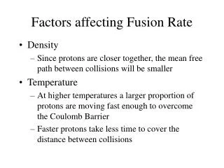
Factors Affecting the Sedimentation Rate. Elevating the ESR: Inflammatory Diseases Cytokine driven processes that elevate fibrinogen ie TB, Pneumonia, rheumatoid arthritis

E N D
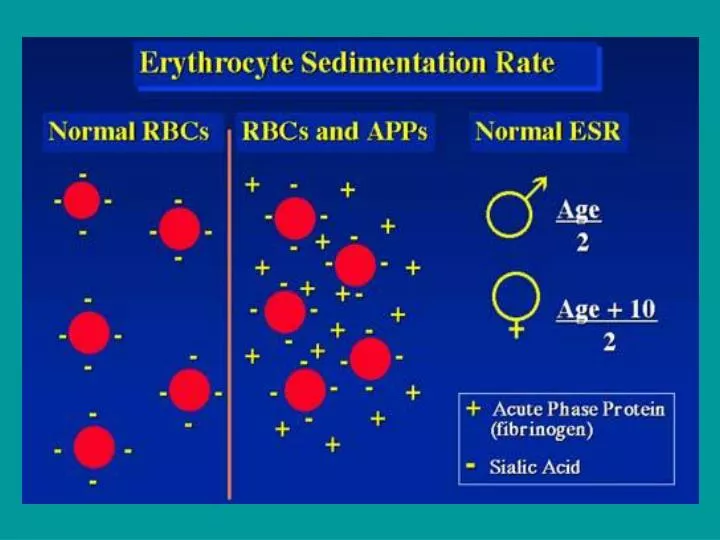
Factors Affecting the Sedimentation Rate Elevating the ESR: Inflammatory DiseasesCytokine driven processes that elevate fibrinogen ie TB, Pneumonia, rheumatoid arthritis • Relative/Absolute Increase in Globulin ProteinsLoss of albumin ie nephrotic syndrome or increase in globulins ie multiple myeloma • Extensive Tissue NecrosisMyocardial infarction, trauma, tumors • Other CausesPregnancy (increase in fibrinogen, anemia), anemia, age, heparinized blood • Lowering the ESR: • Increased Plasma ViscosityWaldenstrom's macroglobulinemia • Red Cell Number or ShapePolycythemia vera, sickle cell disease • Decreased Plasma ProteinsHepatic necrosis, hypofibrinogenemia • Others CausesTrichinosis
Range - The range of normal SRE is 0-15 mm/hr for men and 0-20 mm/hr for women. Many articles have detailed the elevation of the ESR with age and some have suggested the formula of age divided by 2 for men and age plus 10 divided by 2 for women although this has not been universally adopted. • Utility - Although still widely used, the sedimentation rate has limited use as a diagnostic test. It is useful for predicting prognosis in diseases such as rheumatoid arthritis and Hodgkin's disease and it has utility as a marker of treatment efficacy in many diseases such as rheumatoid arthritis, the vasculitides, collagen vascular diseases, and septic arthritis.
CRP (Proteina C reattiva) • The C-reactive protein owes its name to the ability of this protein to precipitate pneumococcal C-polysaccharide in the presence of calcium. It was first discovered in 1930 by Tillet and Frances. • a positive CRP may indicate any of a number of things: • Rheumatoid arthritis • Rheumatic fever • Cancer • Tubercolosis • Pneumococcal pneumonia • Myocardial infartion • Systemic Lupus Erithematosus • Positive CRP results also occur during the last half of pregnancy or with the use of oral contraceptives.
CRP Utility - Because the CRP is a direct measure of inflammation and it is becoming easier and cheaper to do, there may be a time the CRP supersedes the ESR (although the same was said for the measure of plasma viscosity 10 years ago). It is as useful as the ESR in most cases and more accurately reflects the current level of inflammation.
PRINCIPALI MALATTIE AUTOIMMUNI SISTEMICHE INTERMEDIE ORGANO-SPECIFICHE T. HASHIMOTO LES S. GOODPASTURE ADDISON IDIOPATICO AR UVEITE FACOGENICA LUPUS DISCOIDE GASTRITE ATROFICA CIRROSI B. PRIM. DIABETE GIOVANILE OFTALMIA SIMPATICA SCLERODERMIA SCLEROSI MULTIPLA IFERTILITA' MASCH. D. MIOSITE M. DI SJOGREN S. PLURIENDOCRINE AUTOIMMUNI CONNETTIVITE MISTA MIASTENIA GRAVE COLITE ULCEROSA ANEMIA PERNICIOSA
Lupus eritematoso sistematico • Appare nelle donne tra 13 e 40 anni. Rapporto maschio-femmina 1:10 • Caratterizzato da febbre, debolezza, artriti, disfunsioni renali • I pazienti producono autoanticorpi verso il DNA, istoni, eritrociti, piastrine, leucociti, e fattori di coagulazione del sangue • Gli immunocomplessi depositati lungo le pareti dei vasi sanguigni causano una ipersensibilità di tipo III, originano danno endoteliale che da luogo alle reazioni infiammatorie che generano vasculuiti e glomerulonefriti
Esami di Routine e Lupus Incremento della VES e dei livelli di PCR Emocromo: Anemia (Sintomo Costante) Leucopenia (Linfocitopenia) Trombocitopenia Alterazioni della coagulazione (Lupus Anticoagulante) Esame Urine: Albuminuria (Microalbuminuria) Ematuria
Allo stato attuale la dimostrazione della positività della ricerca di autoanticorpi anti-nucleo (ANA) o la presenza di anticorpi anti-dsDNA o anti-Sm (uno degli antigeni nucleari estraibili, ENA) costituiscono 2 degli 11 criteri utilizzati da anni per la diagnosi di lupus eritematoso sistemico
In generale il protocollo diagnostico iniziale, in pazientisintomatici prevede la rilevazione degli anticorpi anti-nucleo in IFI; il pattern di fluorescenza nucleare o citoplasmatico determina la scelta successiva, rappresentata dalla ricerca di autoanticorpi diretti verso uno o più specifici autoantigeni intracellulari
Autoimmunity UV light
Determinazione degli ANA: Tecnica IFI Standardizzata con linee cellulari epiteliali (HEp-2) (esprimono antigeni umani presenti in tutte le fasi del ciclo cellulare) Ai fini diagnostici I titoli di 1:40 e di 1:160 sono considerati come livelli decisionali: Titolo soglia 1:40 (alta sensibilità/bassa specificità) <1:40 negativo. (Anticorpi antinucleo a basso titolo 1:40 - 1:80 possono essere presenti in soggetti sani, nelle gravide, in donne sopra i 40 anni, negli anziani) >1:40 e <1:160 basso positivo (in assenza di sintomi specifici, il protocollo diagnostico deve prevedere un monitoraggio in tempi successivi) >/=1:160 sono da considerare comunque suggestivi di patologia autoimmune.
Pattern omogeneo Fluorescenza omogeneamente diffusa a tutto il nucleo con colorazione dei cromosomi delle cellule in mitosi. Gli Ab sono diretti contro desossiribonucleoproteine, istoni, dsDNA. Pattern periferico Forte fluorescenza alla periferia del nucleo debole al centro. Dovuta alla presenza di autoanticorpi diretti contro ds DNA o contro desossiribonucleoproteine
IL test IFI viene utilizzato anche per la valutazione degli anticorpi anti ds DNA questo test si fonda su una reazione a carico del DNA mitocondriale a doppia elica contenuto nel cinetoplasto di un emoflagellato non patogeno per l’uomo (Crithidia luciliae) Negativo Positivo
Limitazioni di quest’approccio: • Alcuni ENA (anti-Ro/SSA ed anti-Jo1) possono dare risultati falsamente negativi (ridotta espressione degli antigeni-bersaglio nelle cellule HEp-2; perdita e/o denaturazione degli antigeni in fase di allestimento dei vetrini • Non univocità della modalità di refertazione • Dipendenza dell’affidabilità del dato dall’esperienza del microscopista • Difficoltà di reperimento di sieri standard di riferimento
Gli anticorpi anti-DNA • Test EIA • DNA a singola elica (denaturato, ssDNA; determinanti antigenici: zone ricche di G-C e A-T) • DNA nativo a doppia elica (dsDNA, epitopi localizzati lungo lo scheletro glico-fosfato) • Metodica IFI (Crithidia luciliae), • DNA nativo a doppia elica • Gli anticorpi anti-ssDNA non hanno una buona associazione con precisi quadri patologici. Gli anticorpi anti-dsDNA sono altamente specifici per il LES (10° criterio diagnostico del LES)
AutoantigeniENA: • Ro/SS-A • La/SS-B • Sm • RNP • Topoisomerasi I (Scl-70) • Istidil-tRNA sintetasi (Jo-1) • Proteina B centromerica (CENP-B) • rRNP • Nucleosomi (cromatina) • In presenza di segni clinici di S. di Sjogren o di Dermatomiosite/polimiosite possono • rappresentare il principale dato di laboratorio.
Protocolli diagnostici in alcune malattie Autoimmuni • LUPUS • ANA Pattern omogeneo o periferico ad alto titolo • Anti dsDNA Positivo • ENA anti-Sm(altri inutili) • AnticorpiAntifosfolipidi Incostantemente presenti • e lupus Anticoagulante
Protocolli diagnostici di base in alcune malattie Autoimmuni • Sindrome di Sjogren • IgG Incremento policlonale • ANA 20% dei casi negativi • 80% dei casi positivi • 80% granulare • 20% omogeneo • Raramente Nucleolare • Anti dsDNA (Inutili) • ENA 80% Ssa/Ro • 70% SSb/La
Protocolli diagnostici di base in alcune malattie Autoimmuni • Polimiosite/Dermatomiosite • VES Molto Elevata • Emocromo Lieve anemia, Eosinofilia • ANA Pattern granulare • Anti dsDNA (Inutili) • ENA 15% dei casi positivo Jo1 • CPK, LDH Generalmente aumentate • Fattore Reumatoide 30% dei casi positivo
Antiphospholipid antibodies: • Can be present in 30% 0f SLE patients. • Antibodies directed against phosphorylatedpolysaccharide esters of fatty acids • Include: lupus anticoagulant, b2-glycoprotein-I, anti-prothrombin Abs, and anticardiolipin Ab. • False positive VDRL can be seen in 50% of patients • aPL production can also be associated with: Medications Infections Neoplasms (lymphoma)
ANCA: • Abs directed against several proteins in cytoplasm of neutrophils in sera of patients with different Vascular Autoimmune disease and Systemic Autoimmune Disease . • Measured by indirect immunofluorescence. • ELISA is used to detect specific Abs to proteinase-3 , and myeloperoxidase (MPO)
ANCA Patterns: • Cytoplasmic ANCA (C-ANCA): anti-proteinase -3 • Perinuclear ANCA (p-ANCA): anti- myeloperoxidase (MPO), but also elastase and other proteins in the neutrophil granules • Atypical Patterns: Ab to elastase, cathepsin G, lactoferrin, etc.
Gli ANCA: patologie associate Sono considerati i principali markers sierologici specifici delle vasculiti e delle malattie infiammatorie croniche,con un’incidenza dell’85-90% nelle prime e 20-70% nelle seconde ; Vasculite : infiammazione e Malattie infiammatorie croniche necrosi dei vasi con conseguente intestinali (MICI): modificazione del lume vasale ed con questo termine si indicano : alterazioni ischemiche dei tessuti - Rettocolice ulcerosa: irrorati; infiammazione del colon; quella maggiormente - Morbo di Crohn : distruzione rappresentata è il Morbo di Wegener. continua della parete dell’intestino,specialmente a carico del tenue.
Classificazione p-ANCA o ANCA perinucleari : reagiscono principalmente con la mieloperossidasi (MPO) presente nei granuli α-azzurrofili, dando una fluorescenza di tipo perinucleare ; c-ANCA o ANCA citoplasmatici : diretti contro la proteinasi3 (PR3), dando una fluorescenza finemente granulare e diffusa per il citoplasma ; x-ANCA o ANCA atipici : - danno una fluorescenza sia citoplasmatica che nucleare - si osservano nei soggetti affetti da MICI,ma hanno una sensibilità sconosciuta e una specificità bassa.
C-ANCA (cytoplamsic-ANCA): • Diffuse staining of neutrophil cytoplasm in immunofluorescene. • Recognizes PR3(Proteinase –3), a serine protease in primary granules of neutrophils. • Is seen in 85% (range 30-90%) of patients with Wegeners Granulomatosis. • Highly specific for WG (98%) • Titer correlates with disease activity (in 60% of cases) and severity.
P-ANCA: • P-ANCA in the setting of vasculitis is Ab against MPO. • Vasculitidies associated with MPO include: • Microscopic Polyangiitis (45-80%) • Idiopathic Crescentic GN: (65%) • Churg Strauss: (60%) • PAN: (15%) • Anti- MPO can also be due to medications: PTU, hydralazine, minocycline, D-penicillamine. • Atypical P-ANCA can be seen with: RA, SLE, IBD, Chronic liver diseases.
Rheumatoid Arthritis Laboratory Testing, Principals and Guidelines
Rheumatoid Factor (RF)Introduction: • RF is an antibody directed against the FC portion of IgG. • RF was originally described by Waaler and Rose in 1940 • The RF measured in laboratories is IgM RF, but IgG & IgA RF have been described.
Conditions Associated with Positive RF: • Healthy Individuals: • RF can be positive in up to 4% of young and healthy individuals. • RF positivity is higher in elderly people without rheumatic diseases (ranging from 3-25% in different studies). • RF titer in this setting is usually < 1:160
Conditions Associated with Positive RF: …cont. II. Rheumatic Disorders: • Rheumatoid Arthritis: 26-90%* • Sjogren’s syndrome: 75-95% • SLE: 15-35% • Polymyositis/Dermatomyositis:5-10% * Variable in different studies, depending on severity of disease in the study population
Anti-Cyclic Citrullinated Peptide: • Target amino acid is citrulline, in filaggrin molecule derived from human skin. • Citrulline is a post-translationally modified arginine residue. • ELISA assay for anti-CCP may be useful in early stages of polyarthritis. [1] Sensitivity Specificity Anti-CCP 56% 90% IgM RF 73% 82% RF & anti-CCP 48% 96% [1] Bas et al. Rheumatoloy (Oxford) 2003; 62: 870
Serum Electrophoresis • Gel electrophoresis shows an increase in the globulins especially gamma (antibodies) and alpha-2-globulin • There is often a decrease in albumin (7)
Complete Blood Count (CBC) • WBC in the peripheral blood often remained undisturbed • RBC’s show a moderate normocytic hypochromic anemia of chronic disease • A decrease in serum iron is common, Total Iron Binding Capacity (TIBC) and normal iron stores (ferritin) are essentially normal
Creatinine Kinase (CK) • Serum CK is decreased below normal in >60% of RA patients. This is not to be confused with the CKMB (Myocardial) portion or CKMM (skeletal muscle) enzymes that are often measured during a MI or chronic muscular diseases (7)
Synovial Biopsy • Synovial fluid has high WBC and low viscosity, glucose is greatly diminished in the synovium • RF usually present • Characteristic rheumatoid nodules to aide in diagnosis • Positive biopsy of subcutaneous rheumatoid nodule and synovia
American Rheumatism Association Diagnosis Criteria Positive biopsy within diagnostic guidelines: criteria consists of: • Poor mucin clotting of synovial fluid (4) • Characteristic histological changes in synovium: mesothelial, macrophage, LE cells
Testing non-indicative of RA • Other routine serological quantitative testing is normal. This group includes Calcium, Uric Acid, Alkaline Phosphorus, Phosphorus, and Antistreptolysin O titers
Diagnostica di laboratorio nello studio delle patologie immunoallergiche
La diagnostica dell’ asma bronchiale è costituita da: • Anamnesi ed esame obiettivo • Test specifici in vivo e in vitro
VALUTAZIONE ANAMNESTICA • Predisposizione familiare (atopia) o altre patologie immunologiche; • Attività lavorativa • Abitudini di vita (sport, fumo, ambiente) • Terapie in atto • Stagionalitàdegli eventi allergici e modalità di insorgenza
Le reazioni di ipersensibilità IgE mediate determinano diversi tipi sintomatologie L’allergia costituisce l’aspetto patologico, che si traduce in danno,con quadri clinici diversi secondo gli organi interessati: • Rino-congiuntivite • Asma • Orticaria • Allergie intestinali • Shock anafilattico
I TEST UTILIZZATI SONO: IN VIVO Test cutanei PRICK E PATCH TEST IN VITRO PRIST o IgE totali RAST o IgE specifiche
DOSAGGIO IgE TOTALI IL RISCONTRO DI ELEVATI LIVELLI DI IgE TOTALI NON AUTORIZZA UNA DIAGNOSI DI ALLERGIA, POICHE’ POSSONO ESSERE ANCHE PRESENTI IN: • Soggetti normali, • Patologie Parassitarie • Connettiviti, • Infezioni batteriche croniche • Malattie Linfoproliferative
Sistema di classificazione e cut-off relativi alla classe di IgE Specifiche • Elaborati in base alla curva standard e alla calibrazione, forniti in kU/L Classe kU/L 0 <0,35 Assente o non rilevabile I 0,35-0,69 Basso II 0,70-3,49 Moderato III 3,50- 17,49 Elevato IV 17,5-52,49 Molto elevato V 52,5-99,99 Molto elevato VI >100 Molto elevato
EOSINOFILI BASOFILO - Mast cellule T-LINFOCITI EOS IL-x T ISTAMINA PGs - LTs TRIPTASI ECP PBM CHEMOCHINE Basophil DOSAGGIO MEDIATORI E CITOCHINE
ANA: If the ANA is positive, the laboratory automatically performs Extractable Nuclear Antigens (ENA) and Double Stranded DNA Antibody (dsDNA) tests. If the ANA is negative, one may still request the Autoimmune Screen Panel II (Advanced) depending on the working diagnosis. Once the ANA has tested positive there is no diagnostic benefit in repeating this test. Please remember that an ANA may also be positive in up to 5% of normal young females and decisions to proceed with further testing should always take into account overall clinical and laboratory features.