Download
1 / 41
430 likes | 1.23k Vues
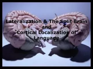
Association Cortex, Asymmetries, and Cortical Localization of Affective and Cognitive Functions. Michael E. Goldberg, M.D. The origins of localization. The concept that different parts of the brain did different things started with Spurzheim and Gall, whose phrenology became quite fashionable:

E N D
Association Cortex, Asymmetries, and Cortical Localization of Affective and Cognitive Functions Michael E. Goldberg, M.D.
The origins of localization • The concept that different parts of the brain did different things started with Spurzheim and Gall, whose phrenology became quite fashionable: • The phrenologist said that a given area of the brain increases in size, as does the overlying skull, when its function is exercised, and a good clinician can, by laying on hands, tell you what parts have been most exercised.
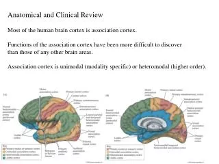
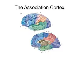
Unimodal cortices Somatosensory/motor Visual Auditory
Association Cortex • Has functions more complicated than simple input and output. • Combines signals from primary sensory and motor modalities to create emergent psychological properties such as • Memory • Planning • Spatial analysis • Language and reading – language associates arbitrary auditory, visual, or tactile symbols with concrete or abstract objects and actions. • Emotion and appetite
Different association cortices have different functions Behavioral Planning Attention Declarative Memory Parietal lobe Frontal lobe Working Memory Spatial location Emotional Processing Response Inhibition Body Image Affective Processing Receptive language Temporal lobe Expressive Language Transfer of sensory information to the motor system
Functions of frontal association cortex • Motor planning – remember the anti-saccade from my oculomotor lecture. That you are here today is largely the responsibility of your frontal cortex. • Working memory. • Suppression of stimulus-bound behavior. • Babies and demented people cannot suppress the urge to urinate in response to a signal from a full bladder • Luckily, you can! • Frontal functions must be studied with complicated paradigms – they are deficits beyond simple sensory failure or motor paralysis • The delayed response task is a paradigmatic task useful in frontal function.
Fixation Sample Delay Test (500-1000 ms) (500 ms) (1000-1500 ms) (<500 ms) Delayed response tasks Matching Non-matching
F C D T Frontal saccade-planning neuron
The neuron is tuned for a specific direction of movement Delay activity (sp./s) Sample location (deg.)
The neuron is tuned for a specific direction of movement not to make. Delay activity (sp./s) Sample location
Functions of prefrontal cortex: • Working memory. • Planning of behavior over long periods of time. • Response inhibition – behaving appropriately. • Complex problem solving, e.g. the Wisconsin card sort task. • Expressive aspects of language
Frontal signs at the bedside • Emergence of primitive reflexes that grownups suppress: • Grasp reflex • Suck reflex • Root reflex • Failure to suppress inappropriate responses to sensory stimuli • Antisaccade • Failure to suppress the blink response to a glabellar tap
Psychiatric aspects of frontal function • Schizophrenics have depressed frontal function by PET and fMRI criteria. • Some schizophrenics and their first order-relatives do poorly on tasks designed to examine frontal function. • Patients with left frontal strokes have a higher frequency of depression than patients with posterior strokes.
Different association cortices have different functions Attention Parietal lobe Spatial location –Where things are Body Image Transfer of sensory information to the motor system
Attention and the parietal cortex • Parietal neurons respond to salient objects in the visual field, not all objects. • Objects can be made salient from bottom-up or top down criteria. • Parietal neurons respond more intensely to attended objects than to unattended objects. • Patients with right parietal lesions neglect the left half of objects and space.
Patients with right parietal lesions neglect the left half of objects and of space
The accurate representation of space • Helmholtz postulated that the brain created a spatially accurate representation of space by associating visual information with a description of the motor signal that moved the eyes – the sense of effort or corollary discharge. • Parietal neurons combine visual and corollary signals to calculate a spatially accurate visual representation.
Parietal visual neurons, like all classic visual neurons, have receptive fields relative to the center of gaze.
RF RF RF FP FP A V V H Stim Start of saccade Start of Saccade Parietal neurons remap their receptive fields around the time of every saccade. RF A H
The parietal cortex sends spatially accurate visual information to the premotor cortex, so accurate movement signals can be generated. • Where objects are in space. • How big they are. • What is their orientation.
Parietal signs at the bedside - apraxia • Apraxia – inability to conceptualize or mimic a movement, even though the patient can make the necessary movements – patients with parietal lesions cannot mimic how to use a toothbrush but they can use one. They cannot orient their hands or set a grip in a movement. • Constructional apraxia - they cannot duplicate block designs, and have great difficulty copying drawings. • Optic ataxia – difficulty reaching to objects in space or finding them with saccades.
Parietal signs at the bedside – attentional and body-image deficits • Extinction – neglect of a stimulus in the affected field – visual, tactile, or auditory – when presented simultaneously with an equivalent stimulus in the normal field. • Anosognosia – patients do not recognize the contralateral (usually left) limb as a part of their own body. • Spatial distortion.
Different association cortices have different functions Declarative Memory Emotional Processing Receptive language Temporal lobe
Temporal and Limbic Cortex • Hippocampus – declarative memory. • Amygdala – emotional processing and fear (not really cortex, but deep in the temporal lobe). • Rhinal cortex – associating motivational value to visual objects. • Temporal neocortex is mostly unimodal – auditory and visual. • Wernicke’s area for expressive aphasia lies at the border of the temporal and parietal lobes.
H.M. – Rasumussen and Milner’s patient with a bilateral hippocampal excision for intractable epilepsy.
H.M.’s deficits • He could not consciously remember any fact for more than about 45 sec. Brenda Milner examined him almost every day for years, and he never recognized her. • He could learn motor skills such as tracing a maze, which required practice. • His epilepsy was much improved
Temporal signs at the bedside • Receptive (Wernicke’s) aphasia. • Korsakoff’s syndrome – requires bilateral destruction of the output of the hippocampus – fornix and mammilary body. • Temporal deficits are more often behavioral: difficulty relating to others, sexual problems, emotional problems. • The damaged hippocampus often evokes seizures that start with complex auras and produce complex behavioral automatisms.
Hemispheric asymmetry • Dominance refers to the hemisphere with speech – usually left hemisphere. • In left-dominant subjects the right hemisphere does more spatial analysis. • Children who have strokes in their dominant hemisphere before the age of 2 develop normal speech, but lose some spatial ability as judged by psychometric spatial tasks.
Primary sensory modalities are contralateral. Information from one hemi visual field or one side of the body reaches the ipsilateral cortex through the corpus callosum. Patients with severe epilepsy can sometimes be helped by section of the corpus callosum. Interhemispheric communication
Patients with callosal section • Have their entire right hemisphere disconnected from the speech area • Stimuli in the left visual field and the left side of the body only go to the right hemisphere. • Stimuli in the right visual field and right side of the body only go to the left hemisphere. • The left hemisphere does not know about, and cannot talk about, information limited to the right hemisphere.
Callosal section and reading Sex Neurobiology is Cool! (symbols) Neurobiology is Cool! (semantic meaning) Giggle ???????? Neurobiology is Cool! Sex
Alexia without agraphia, a callosal disconnection syndrome • Patients have a lesion of the left visual cortex and the splenium (most posterior part) of the corpus callosum. • Visual information cannot get to the speech area, so the patients cannot read. • Visual information can get to the motor area, so they can write. • They can’t read what they have written. • They can’t name colors, although they can match colors.
Alexia without agraphia Neurobiology is Cool! Neurobiology is Cool! ???????? Neurobiology is Cool!
Take home message • Association cortex combines information from multiple modalities – sensory, motor, emotional. • Frontal association cortex plans behavior and facilitates working memory. • Parietal association cortex analyzes space, generates attention, and transmits sensory information to the motor system. • Temporal cortex (hippocampus) organizes declarative memory.
More take home message • Speech is mostly located in the left hemisphere. • Spatial processing is mostly located in the right hemisphere. • The corpus callosum connects the two. • Damage to the corpus callosum prevents interhemispheric communication.
More errata in KSJ(not my fault this time) • Posterior parietal cortex (area 7) is an association area with visual, auditory, and somatosensory, and motor corollary inputs. • The temporo-parietal polysensory area is another area of multimodal associations about which less is currently known.