Download
1 / 41
410 likes | 978 Vues
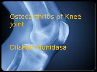
OSTEOARTHRITIS: (OA) OSTEOCHONDROSIS. OSTEOARTHRITIS: (OA) - It's a degenerative process involving the whole joint, but beginning in the articular cartilage. Other features. Causes of osteoarthritis Excessive load

E N D
OSTEOARTHRITIS: (OA) - It's a degenerative process involving the whole joint, but beginning in the articular cartilage.
Causes of osteoarthritis Excessive load • The total force crossing a joint may be excessive or applied over too small an area, or both factors may be combined. • Total force-too great. It is not generally realized how great are the forces to which articular cartilage is subjected. Thus, in a normal hip the lever system is such that with each step a force of 4 to 6 times the body weight is transmitted. If the lever system is faulty, as in Coxa Valga, the force is greater still. Similarly with genu varum the force increases very considerably with each centimeter that the knee deviates from the center of gravity of the body.Objects being carried (including surplus fat) obviously increase these forces still further.
Area too small. With joint deformity the area available for load transmission is usually reduced. Obvious examples are congential hip subluxation(in which part of the head is lateral to the acetabulum and an old slipped upperfemoral epiphysis. • Both factors combined. In genu varum, congential hip subluxations, andsimilar disorders, not only is the area for load transmission too small, but theactual force also is increased because of the faulty lever system.
Defective cartilage. • The articular cartilage may be injured, affected by disease, or deprived of support by normal bone. • 1) Damaged Cartilage. Articular Cartilage may be damaged by a single major injury such as an osteochondral fracture. Repeated minor injury is equally damaging and occurs in a wide variety of condition such as, recurrent subluxation of the Patella. Certain occupations or recreations (for example at the elbow with javelin throwing) undue joint laxity (congential, following ligament injury, or when ligaments and capsule are insensitive to pain or positions), and the repeated abrasive action of loose bodies or a torn meniscus. • In hemophitia repeated bleeding leads to synorthis, hydrolytic enzymes and liberated causing an inflammatory response which damage Cartilage, further damage results from the accumulation of iron within individual cells. A similar effect may account for the joint degeneration in haemochromatosis.
Defective cartilage 2) Diseased Cartilage. The Cartilage may be weakened by inflammatory or metabolic disorders. • Inflammatory disorders: Rheumatid arthritis is the commonest inflammatory cause of weakened articular cartilage, but unrecognized low grade inflammation is probably not uncommon and the associated bone softening might explain the appearance in protrusion acetabulae. Much less causes are septic and tuberculous arthritis. • Metabolic Disorders: Two metabolic disorders, which weaken articular cartilage (gout and pseudogout). A much rarer example is alkoptonuria in which homogertisic acid is deposited in the urine which turns black on standing and sometimes in cartilage which also looks black (hence the term ochronotic arthropathy ), X-ray shows calcified intervertebral discs and menisci.
Defective cartilage 3) Unsupported Cartilage. If subchondral bone is deprived of its blood supply it becomes necrotic (a vascular necrosis). Obvious examples are caisson disease, hip fracture and dislocation.
Symptoms • Pain:This leading symptom. In early osteorthritis pain occurs after a nights rest and then wear off. Later after use this joint aches again. As the disease progress, pain becomes more severe and more constant. Sometimes disturbing sleep. Three distinct varieties of pains have been described, capsular (on forcing extremes), muscular (following exertion) and venous (after standing, and at night). • Stiffness:This too is an important symptom. At first it is noticed only after the joint has been still for some time. Later it is constant and gradually increases.
Symptoms • Deformity. Capsule shrinkage and muscle imbalance produce deformity. At the hip the patient may notice increasing shortness of the leg because of flexion and adduction deformity. • Limp. This is common and due to pain or deformity. • Giving way. This sometimes occurs, possibly because a synovial fringe has been nipped, or occasionally because an osteophyte has broken off from a loose body.
Symptoms • Swelling. Swelling is only noticeable at superficial joints such as the knee. • Signs. The patient is fit. One joint is only affected; occasionally 2 or even 3 may be involved, but this in not a true polyarticular disease like rheumatoid arthritis. The local signs in an affected joint are as follows: • Look. There are no scars only slight wasting. Swelling may be due to the fluid (in the knee) or to osteophytes (at the metatarsophalangeal joint of the ha-luix. Deformity may be obvious the hip may be hold flexed, adducted and externally rotated.
Symptoms • Feel. They're in no warmth. In superficial joints localized tenderness is common, and fluid or osteophytes may be felt. • Move. Movement is always restricted but is painless within the permitted range. The Restriction is characteristically asymmetrical, some movements much more than others. Thus ,at the hip, extension, abduction and internal rotation are far more limited than their opposites. Creptius is common.
Radiographs are normal early in the disease, but narrowing of the joint space develops as the disease progresses. Ninety percent of individuals over the age of 40 years have radiographic changes characteristic of osteoarthritis, however, only 30% have symptoms. Other x-ray features include subchondral sclerosis, marginal osteophytes, and subchondral cysts.37 In osteoarthritis, subchondral cysts are surrounded by a dense rim of bone that differentiates them from the marginal erosions that occur in rheumatoid arthritis. Laboratory features in arthritis are nonspecific and are generally not helpful in making the diagnosis.
Treatment. • There are several ways of treatment of osteoarthritis. The most common is a way of using drugs for killing pain or injections of corticosteroids in the soft tissues, surrounding the bones. Often liniments are used. The second way is physiotherapy, which includes magnetotherapy, ultrasound therapy, hot water baths, message, radiant heat, all methods of applying superficial warmth; short-wave diathermy penetrates more deeply. To this conservative way of treatment we have to include X-ray therapy, which means the use of X-rays on • the joint area. Such a way of treatment provides an anti-inflammatory and analgesic effects. The efficacy is very high — more than 90% of all cases.
Operativetreatment • Arthrodesis: This is the only certain way to abolish pain permanently, but the penalty of total stiffness is to increase the shesses on other joints. • Arthroplasty: Although theoretically it is more desirable, arthroplasty is less certain to relive pains. The join capsule (with it's pain fibers) needs to be removed, what makes such an operation quite traumatic. • Osteotomy: Pain is often relived because shess is now distributed more widely and because the capsule is no longer tight in position of habitual use. Ultimately the joint undergoes remarkable "biological regeneration", again it may be due to better distributions of pressure, but the relief of venous congestions in the subchondral sinusoids may be a factor.
Cervical Spondylosis • Cervical spondylosis is defined as a generalized disease process affecting the entire cervical spine and related to chronic disk degeneration. In approximately 90% of men older than 50 years and 90% of women older than 60 years, degeneration of the cervical spine can be demonstrated by radiographs. Initial disk changes are followed by facet arthropathy, osteophyte formation, and ligamentous instability. Myelopathy, radiculopathy, or both may be seen secondarily. Cervical myelopathy is the most common form of spinal cord dysfunction in people older than 55 years. People older than 60 years are more likely to have multisegmental disease. The incidence of cervical myelopathy is twice as great in men as in women.
Symptoms and Signs • Headache may be the presenting symptom of cervical spondylosis. Usually, the headache is worse in the morning and improves throughout the day. It is commonly located in the occipital region and radiates toward the frontal area. Infrequently, patients complain of a painful, stiff neck. Signs include decreased range of motion, crepitus, or both. With more advanced cases, radicular or myelopathic symptoms may be present.
Cervical Spondylotic Radiculopathy • Cervical radiculopathy in spondylosis can be quite complex, with nerve root involvement seen at one or more levels and occurring either unilaterally or bilaterally. The onset may be acute, subacute, or chronic, and impingement on the nerve roots may be from either osteophytes or disk herniation. With radiculopathy, sensory involvement in the form of paresthesias or hyperesthesia is more common than motor or reflex changes. Several dermatomal levels may be involved, with radiation into the anterior chest and back. The chief complaint is radiation of pain into the interscapular area and into the arm. Typically, patients have proximal arm pain and distal paresthesias.
Imaging Studies • Although spondylosis results from cervical spine degeneration, not every patient with radiographic evidence of cervical disk degeneration has symptoms. Furthermore, patients with all the radiographic stigmas of cervical spondylosis may be asymptomatic, and others with clinical evidence of myelopathy may show only modest radiographic changes. This paradox is explainable by canal size differences, with the smaller-diameter canal having less space to buffer the degenerative lesion. • Plain film findings also vary according to the stage of spondylosis at which they were taken. Radiographs may appear normal in early disk disease. Alternatively, they may show single or multilevel disk space narrowing with or without osteophytes. C5-C6 and C6-C7 are the two most commonly involved segments (Figure 5–7). Vertebral body sclerosis at the adjacent base plates may also be seen. Cortical erosion is uncommon and indicates an inflammatory process such as rheumatoid arthritis.
Treatment • Patients should be divided into three groups, according to the predominance of their symptoms: neck pain alone, radiculopathy, and myelopathy. The duration and progression of symptoms need to be considered in the planning of treatment. Several studies suggest that patients with cervical radiculopathy or myelopathy have better long-term results from surgery if symptoms are of short duration.
Conservative Treatment • Initial management of patients with cervical spondylosis may involve a soft collar, antiinflammatory agents, and physical therapy consisting of mild traction and the use of isometric strengthening and range-of-motion exercises. The soft cervical collar should be worn only briefly, until the acute symptoms subside. Analgesics are important in the acute phase, and muscle relaxants are helpful in breaking the cycle of muscle spasm and pain. Diazepam should be avoided because of its side effects as a clinical depressant. Epidural corticosteroid injections may be efficacious in patients with radicular pain. Trigger point injections are an empirical form of therapy that seems to work well in patients with chronic neck pain. • The value of cervical traction remains unclear. It is contraindicated in patients with cord compression, rheumatoid arthritis, infection, or osteoporosis. A careful screening of roentgenograms before treatment is mandatory. No evidence indicates that home traction is more effective than manual traction. Isometric strengthening exercises of the paravertebral musculature should be started after the acute symptoms resolve. The patient should be instructed to start a home exercise program early, to avoid long-term dependency on passive therapy modalities. Although ice, moist heat, ultrasound, and transcutaneous electrical nerve stimulation (TENS) are safe to use, there is no scientific proof of their efficacy.
Surgical Treatment • Surgical intervention should be considered if the patient does not respond to a conservative treatment protocol or shows evidence of deteriorating myelopathy or radiculopathy. The spinal cord can be effectively decompressed by either anterior, posterior, or combined approaches. • The anterior approach allows multilevel diskectomy, vertebrectomy, foraminotomy, and fusion with tricortical iliac crest bone grafts or strut grafts. Newer instrumentation techniques, such as cervical plates (Figure 5–8), alleviate the need for halo immobilization. However supplemental posterior fixation and fusion should be added if more than three vertebral levels are decompressed anteriorly. Posterior fixation minimizes the risk of anterior dislodgement of the graft even in the presence of solid anterior fixation. Anterior interbody fusion after decompression for a herniated cervical disk (Figure 5–9) has a high success rate.
A: Radiograph showing degenerative changes between C4 and C7. B and C: Radiographs taken after anterior vertebrectomy of C5 and C6, iliac crest strut graft, and anterior plate fixation.
Ankylosing Spondylitis Ankylosing spondylitis is a chronic seronegative inflammatory disease that affects the axial skeleton, especially the sacroiliac joints, hip joints, and spine. Extraskeletal involvement is found in the aorta, lung, and uvea. The incidence of ankylosing spondylitis is 0.5–1 per 1000 people. Although males are affected more frequently than females, mild courses of ankylosing spondylitis are more common in the latter.
Symptoms and Signs The onset is insidious, with early symptoms including pain in the buttocks, heels, and lower back. Patients complain typically of morning stiffness, the improvement of symptoms with activity during the day, and the return of symptoms in the evening. The earliest changes involve the sacroiliac joints and then extend upward into the spine. Spinal disease results in loss of motion and subsequent loss of lordosis in the cervical and lumbar spine. Synovitis in the early stages leads to progressive fibrosis and ankylosis of the joints during the reparative phase. Enthesitis occurs at the insertion of the annulus fibrosus on the vertebral body with eventual calcification that results in the characteristic "bamboo spine." The pain from the inflammatory process subsides after full ankylosis of the affected joints occurs. Approximately 30% of patients develop uveitis, and 30% have chest tightness. Limited chest expansion indicates thoracic involvement. Fewer than 5% of patients have involvement of the aorta, characterized by dilation and possible conduction defects. In addition, patients may suffer from renal amyloidosis and pulmonary fibrosis.
The earliest radiographic changes are visible in the sacroiliac joints. Symmetric bilateral widening of the joint space is followed by subchondral erosions and ankylosis. Bony changes in the spine affect the vertebral body. Changes include loss of the anterior concavity of the vertebral body, squaring of the vertebra, and marginal syndesmophyte formation, which give the spine the appearance of bamboo. Ankylosis of the apophyseal joints also develops. The disease generally starts in the lumbar spine and migrates cephalad to the cervical spine. Atlantoaxial instability is seen occasionally.
Treatment • The natural history of ankylosing spondylitis, with its slow progression over several decades, has to be considered in planning treatment. Initially, treatment consists of exercises and indomethacin. Approximately 10% of patients develop severe bony changes that eventually require surgical intervention. These changes characteristically include a fixed bony flexion deformity that limits their ambulatory potential. Hip disease should be addressed before correction of spinal deformities because correction of hip flexion deformities may allow significant compensation of the spinal kyphosis to allow adequate horizontal gaze. • Loss of lumbar lordosis can be treated by multilevel V-shaped osteotomies posteriorly (the Smith-Petersen procedure), by a decancellation procedure (the Heinig procedure) of L3 or L4, or by pedicle subtraction osteotomy based at L3 or L4.