Download
1 / 58
720 likes | 1.29k Vues
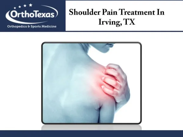
Shoulder Pain. Shoulder Anatomy: Bony Anatomy. Humerus Scapula Glenoid Acromion Coracoid Scapular body Clavicle Sternum. Shoulder Anatomy: Joints. Glenohumeral Acromioclavicular. Sternoclavicular Scapulothoracic articulation. Glenohumeral Joint. Most common dislocated joint

E N D
Shoulder Anatomy:Bony Anatomy • Humerus • Scapula • Glenoid • Acromion • Coracoid • Scapular body • Clavicle • Sternum
Shoulder Anatomy:Joints • Glenohumeral • Acromioclavicular • Sternoclavicular • Scapulothoracic articulation
Glenohumeral Joint • Most common dislocated joint • Lacks bony stability • Composed of: • Fibrous capsule • Ligaments • Surrounding muscles • Glenoid labrum
Shoulder Anatomy:Rotator Cuff Muscles • Depress humeral head against glenoid
Shoulder anatomy:Rotator cuff muscles • Supraspinatus: • Abduction • Infraspinatus: • External rotation • Teres Minor: • External rotation • Subscapularis: • Internal rotation
The supraspinatustendon is the most studied tendon of the rotator cuff because it is the most commonly affected tendon in rotator cuff pathology. The supraspinatus tendon is relatively thick as it courses between the superior aspect of the humeral head and the inferior aspect of the acromion.
Normally, a well-defined space exists between the tendon and the acromion: thesubacromial space. This space is occupied by the subacromial bursa. Laterally, the space and the bursa generally extend beyond the lateral aspect of the acromion underneath the deltoid muscle .
Shoulder Anatomy:Other Musculature • Pectoralis major, deltoid, latissimus dorsi, biceps • Rhomboids, trapezius, levator scapulae, serratus anterior
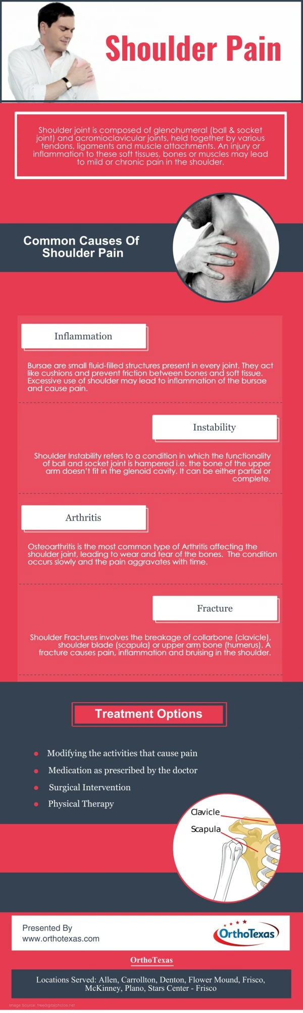
The causes of shoulder pain are numerous. • Acute injuries of the shoulder are often obvious. Shoulder fractures, dislocations are among the most frequent acute shoulder injuries. II. Chronic shoulder pain often has a less obvious cause. Common causes include: • Adhesive Capsulitis • chronic glenohumeral instability, • rotator cuff tendon pathology, • degenerative changes in the acromioclavicular joint • labral pathology
History • Mechanism of injury • Specific sport/activity when injury occurred • Duration of symptoms • Acute event or chronic • Aggravating/alleviating factors • Pain (Location/Character/Night pain) • Sensation of instability
Weakness • Popping/Crepitus: painful/non-painful • Stiffness • Numbness/Tingling • Shoulder activities involved in patients occupationPast medical history of shoulder injury/surgery • Previous history of injections • Hand dominance
Physical Examination • Inspection • Palpation • Range of Motion • Strength • Provocative shoulder testing • Neurovascular status • Neck & elbow exam
Inspection • Compare to normal shoulder for obvious deformities • Abnormalities of: • Humeral head • Clavicle • AC joint • SC joint
Inspection • Muscle atrophy • May indicate nerve damage or disuse atrophy 2° to rotator cuff pain/tear • Appearance of skin: • Swelling • Ecchymosis • Erythema • Venous distention
Inspection • Scapulothoracic motion • Dyskinesia or winging
Palpation • Bony structures: • SC joint • Clavicle • AC joint • Acromion • Greater tuberosity • Coracoid process • Spine of scapula • Soft tissue structures • Short & long heads of biceps • Subacromial bursa • Musculature of shoulder • Anterior capsule • Posterior capsule • Pericapsular musculature
Range of Motion • Passive & Active • Compare to unaffected side • Pain w/ movement? • Dominant shoulder (“Overhead athletes”) • 5° to 10° more external rotation • 5° to 10° less internal rotation
Range of Motion • Forward Flexion • Abduction • Adduction • Internal Rotation • External Rotation
Muscle testing • Compare to unaffected side • Differentiate between true weakness & weakness due to pain
Muscle TestingSupraspinatus • Empty Can Test • 90° abduction • 30° forward flexion • Thumbs pointing downward • Patient attempts elevation against examiner’s resistance
PE: Muscle testingSubscapularis • “Lift-off test” • Internally rotate shoulder • Dorsum of hand against lower back • Patient attempts to push away examiner’s hand • Modified: Place hand on abdomen and resist internal rotation
PE: Muscle TestingInfraspinatus/Teres Minor • Patient’s arms adducted @ sides • Elbows flexed to 90° • Patient attempts external rotation against examiner’s resistance
Provocative Tests • Impingement signs • AC Joint • Biceps tendon • Glenohumeral joint stability • Labral signs • Cervical spine signs
Impingement Signs:Hawkins’ Test • Patient’s arm forward flexed to 90° • Elbow flexed to 90° • Shoulder forcibly internally rotated by examiner • Pain subacromial impingement or rotator cuff tendinitis
Rotator Cuff sign:Drop Arm Test • Passively abduct patient’s shoulder • Observe as patient slowly lowers arm to waist • If arm drops to patient’s side, suggests rotator cuff tear &/or supraspinatus dysfunction
AC joint:Crossover Test • Patient raises affected arm to 90° • Actively adducts arm across body • Forces acromion into distal end of clavicle • Isolates AC joint & painful if positive
Biceps Tendon:Speed’s Test • Elbow flexed 20°-30° • Forearm supinated • Arm in 60° flexion • Patient forward flexes arm against examiner’s resistance
Anterior Instability Testing:Apprehension Test • Supine, sitting or standing • Arm abducted to 90° • Apply slight anterior pressure & slowly externally rotate • Apprehension may indicate anterior instability • Pain w/out apprehension is more likely impingement
Inferior Instability Testing:Sulcus Sign • Arm in neutral position • Pull downward on elbow or wrist • Observe for depression lateral or inferior to acromion • Positive if > 1 cm • Indicates inferior instability • Compare to other side
Posterior Instability Testing:Posterior Apprehension Test • Supine or sitting • Arm in 90° abduction, 90° elbow flexion • Apply posteriorly directed force in attempt to displace humeral head posteriorly
Labral signs • O’Brien’s test • Arm forward flexed to 90° • Elbow fully extended • Arm adducted 10° - 15°, thumb down • Downward pressure • Repeat w/ palm up • Suggestive of labral tear if more pain w/ thumb down
Cervical Spine:Spurling’s Maneuver • Neck extended • Head rotated toward affected shoulder • Axial load placed on the spine • Reproduction of patient’s shoulder/arm pain indicate possible nerve root compression
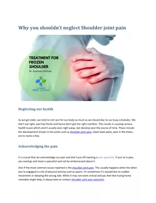
Patients who present with a painful stiff shoulder are frequently diagnosed with “frozen shoulder.” does not denote a specific pathology. Rather, it applies to what he described as “many conditions which cause spasm of the short rotators or adhesions about the joint or bursae.”
Adhesive capsulitisis a specific pathologic entity in which chronic inflammation of the capsule subsynovial layer produces capsular thickening, fibrosis, and adherence of the capsule to itself and to the anatomic neck of the humerus. The contracted, adherent capsule causes pain, especially when it is stretched suddenly, and produces a mechanical restraint to motion.
Many conditions can produce symptoms similar to those of adhesive capsulitis, these include: • full and partial-thickness rotator cuff tears • calcific tendinitis, • glenohumeral or acromioclavicular arthritis • cervical radiculopathy. In these conditions, motion loss is typically multifactorial rather than the result of isolated capsular restriction.
Pathology Adhesive capsulitis is characterized by : • thickened, tight glenohumeral joint capsule • adhesions obliterating the normally patulous axillary fold.
The fibrotic capsule adheres to itself and to the anatomic neck of the humerus. There is minimal synovial fluid in the joint, and overall joint volume is diminished. Normal shoulder joint volumetric capacity is 28 to 35 mL of injected fluid,whereas in adhesive capsulitis, the joint accepts only 5 to 10 mL.
Four stages of disease have been described The disease progresses from capsular inflammation to fibrosis . • Stage 1, the preadhesive stage, consists of a fibrinous inflammatory synovitic reaction without adhesion formation. At this stage, patients typically have full motion but report pain, particularly at night. Symptoms are nonspecific, and misdiagnosis is common.
In stage 4, the chronic stage, adhesions are fully mature, and motion is severely reduced. Patients may have painless, limited range of motion in stage 4, but pain occurs when the arm is suddenly moved beyond the limits of the scarred capsule.
Clinical picture • Most patients with adhesive capsulitis are women aged 40 to 60 years. • The nondominant arm typically is most affected. • Adhesive capsulitis is more common in persons in sedentary vocations than in persons who perform manual labor. • There are numerous association with systemic conditions • cardiovascular disease • thyroid dysfunction, • breast cancer treatment. • Patients with cerebrovascular accident, • myocardial infarction • diabetes are at increased risk of developing adhesive capsulitis. Diabetes is associated with a significantly worse prognosis, greater need for surgery, and suboptimal results.
pain • Loss of motion Patients typically present with pain of insidious onset of several months duration. The onset of symptoms tends to be more gradual than in other shoulder conditions. Pain is commonly referred to the origin of the deltoid. Night pain is common, and patients typically cannot sleep on the affected side.
Loss of motion accompanies or in rare cases precedes the onset of pain and typically becomes more prominent with disease progression. • Patients have difficulty dressing, combing their hair, reaching to a back pocket. • In stages 3 and 4, motion loss becomes severe, affecting movement in all directions. Pain is reduced and may be apparent only when the shoulder is moved beyond the limits allowed by the contracted capsule
Physical examination • does not reveal a specific point of tenderness. • Occasionally, the long head of the biceps tendon is tender because its synovium is confluent with that of the glenohumeral joint. • Rotator cuff strength is usually normal. • A mechanical restraint to passive motion is the hallmark of adhesive capsulitis. This finding is best appreciated on passive external rotation with the arm at the side.
Although radiographs are typically normal, they are important for eliminating other causes, such as arthritis, calcific tendinitis, and unrecognized shoulder dislocation (especially posterior). Disuse osteopenia may be seen in patients with long-standing disease.