Download
1 / 58
611 likes | 1.01k Vues
Gross Brain Overview: Part I. Basic Neuroscience James H. Baños, Ph.D. Overview. Anatomical Directions Cranial Fossa Meninges Pathology of the Cranial Fossa & Meninges. Anatomical Directions. Dorsal fin. Anatomical Directions. Dorsal fin. Anatomical Directions. Dorsal fin.

E N D
Gross Brain Overview: Part I Basic Neuroscience James H. Baños, Ph.D.
Overview • Anatomical Directions • Cranial Fossa • Meninges • Pathology of the Cranial Fossa & Meninges
Anatomical Directions Dorsal fin
Anatomical Directions Dorsal fin
Anatomical Directions Dorsal fin
Anatomical Directions Dorsal Ventral
Anatomical Directions Dorsal Caudal Caudus = tail Rostral Rostrum = Stage or speaking platform Ventral
Anatomical Directions Dorsal-ventral and rostral-caudal are with respect to the neuraxis Dorsal Rostral Caudal Ventral
Anatomical Directions But wait……we evolved
Anatomical Directions But wait……we evolved… Dorsal Caudal Rostral Ventral Dorsal Rostral Caudal Ventral Ventral Dorsal Caudal
Anatomical Directions Rostral Caudal Caudal
Anatomical Directions Dorsal Ventral Dorsal Ventral
Anatomical Directions Anterior-posterior and superior-inferior are in 3-D space And do not respect the neuraxis. Superior Posterior Anterior Superior Inferior Anterior Posterior Inferior
Anatomical Directions Medial-lateral can also respect either the midline, or some other reference point Lateral A B Medial
Anatomical Directions Just to keep it interesting: Mesial = Medial
Anatomical Directions Proximal Distal
Ipsilateral vs. Contralateral • Ipsilateral -- On the same side, with respect to a reference point • Contralateral -- Opposite side, with respect to a reference point
Anatomical Planes Sagittal Plane Horizontal/axial Plane Transverse Section Coronal Plane
Coronal Plane Corona = crown….but think Statue of Liberty
Sagittal Plane Mid-sagittal plane Sagittal MRI
Horizontal Plane • AKA: • Axial plane • CT plane
Cranial Fossa • Distinct areas of the skull base • Contain and support various parts of the brain • Contact point for the meninges
Cranial Fossa Anterior cranial fossa -- frontal lobes Middle cranial fossa -- Temporal lobes Posterior cranial fossa -- Cerebellum and brain stem
Cranial fossa contain lumen, or holes forcranial nerves, arterial supply, and venous drainage
Meninges • Three layers covering the brain • Dura mater • Arachnoid • Pia mater • Support, protection • Contain circulating CSF that supports the brain
Dura Mater • “Tough Mother” • Thick, leathery outer covering • Adheres to inner surface of skull • Support, and forms sinus system (veinous drainage) • Pain sensitive • Has own blood supply -- Middle meningeal artery
Arachnoid • Spongy/fibrous layer between the dura and pia • Collagenous connective tissue • Keep brain suspended in meninges • Subarachnoid space • Contains CSF
Pia Mater • Innermost layer • Adheres almost indistinguishably to cortical surface, including sulci and gyri • Difficult to see grossly or remove
Dura Mater Middle meningeal artery
Pia and arachnoid together are sometimes referred to as the leptomeninges • CSF flows between the arachnoid and pia in the subarachnoid space
Dural Reflections • Also called dural septa • In certain places, the dura doubles on itself and forms a division or partition between parts of the brain • Falx cerebri • Tentorium cerebelli
Falx Cerebri • Extends down the longitudinal fissure, between the cerebral hemispheres • Forms the triangular-shaped superior sagittal sinus superiorly
Tentorium Cerebelli • Extends inward between the cerebellum and occipital lobes • Forms a “soft top” for the posterior cranial fossa • Brain stem passes through tentorial notch • Forms the transverse sinus posteriorly
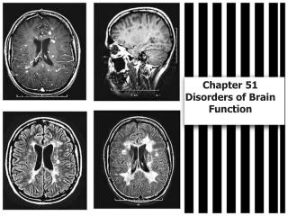
Meningitis • Infection/inflammation of the meninges • Bacterial • Viral • Can progress to • More widespread CNS infection • Hydrocephalus, if flow of CSF is blocked (e.g., purulent accumulation)
Bleeds • Epidural Hematoma • Rupture of middle meningeal artery • Blood accumulates between dura and skull • Mass effect compresses brain • Fed by arterial perfusion…can grow quickly
Bleeds • Epidural Hematoma Mass effect Midline shift
Bleeds • Subdural Hematoma • Rupture of bridging veins from surface of cortex to veinous sinuses (e.g., superior sagittal sinus) • Accumulates more slowly (veins) • Still results in mass effect and compression of the brain • Older people particularly susceptible. Why?