Download
1 / 65
710 likes | 1.09k Vues
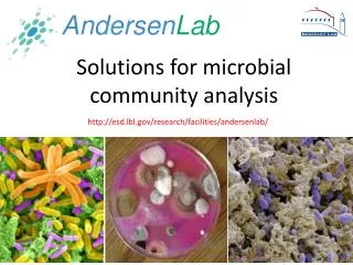
Advances in microbial analysis. Prof. dr. ir. W. Verstraete Dr. ir. N. Boon Laboratory of Microbial Ecology and Technology (LabMET) Faculty of Bioengineering Ghent University LabMET.Ugent.be. Methods to examine microbial populations. Introduction Historical overview Microscopy

E N D
Advances in microbial analysis Prof. dr. ir. W. Verstraete Dr. ir. N. Boon Laboratory of Microbial Ecology and Technology (LabMET) Faculty of Bioengineering Ghent University LabMET.Ugent.be 1Laboratory of Microbial Ecology and Technology
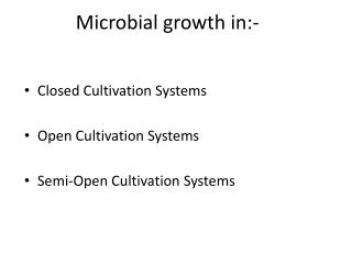
Methods to examine microbial populations • Introduction • Historical overview • Microscopy • Activity measurements • Great Plate Count Anomaly • Immunological methods • Molecular detection methods • PCR detection • Real Time PCR quantification • Microbial fingerprinting • Whole cell analysis • Fluorescent in situ Hybridisation (FISH) • Flow cytometry • Conclusions and perspectives 2Laboratory of Microbial Ecology and Technology
Methods to examine microbial populations • Introduction • Historical overview • Microscopy • Activity measurements • Great Plate Count Anomaly • Immunological methods • Molecular detection methods • PCR detection • Real Time PCR quantification • Microbial fingerprinting • Whole cell analysis • Fluorescent in situ Hybridisation (FISH) • Flow cytometry • Conclusions and perspectives 3Laboratory of Microbial Ecology and Technology
Introduction • What is known about the microbial diversity? 4Laboratory of Microbial Ecology and Technology
Introduction • What kind of information do we want to obtain? • In general: • Diversity: what kind of bacteria are present? • Function: are the essential players present? are there unwanted species? • Practical: • Speed: less than 1 day analysis time (online ?) • Accuracy: be sure of the results • Sensitivity: also the less abundant species... • High-throughput: many samples and many organisms 5Laboratory of Microbial Ecology and Technology
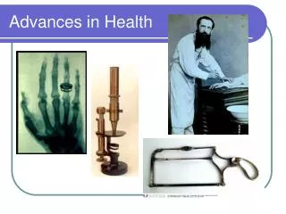
Introduction • Microbial detection: historical overview 6Laboratory of Microbial Ecology and Technology
Introduction • Van Leeuwenhoeks microscope:First observation by a microscopein 1674: “animalcules” 7Laboratory of Microbial Ecology and Technology
Introduction • Microscopy: • Quick • Low specificity Electron microscope (nm) Light microscope (mm-µm) Confocal fluorescence microscope (µm) 8Laboratory of Microbial Ecology and Technology
Introduction • Activity measurements: • Microbial processes: respiration,nitrification, dehydrogenase and phosphatase • Information about bacterial activity • Nothing known about microbial composition 9Laboratory of Microbial Ecology and Technology
Introduction • Koch-1882 and Petri-1887:Culturing micro-organisms on media 10Laboratory of Microbial Ecology and Technology
Sample Plating Counting Introduction • Culturing based methods: • Isolation and enumeration of microbial cells on specific nutrient agars • currently most used approach Results after min 48 h Total Count, coliforms, E. coli, Legionella pneumophila, Clostridia, Salmonella, Aeromonas, ... 11Laboratory of Microbial Ecology and Technology
Introduction The great plate count anomaly (Amann, 1990) 12Laboratory of Microbial Ecology and Technology
Introduction • Limitations of culturing techniques: • The exact growth-conditions are unknown: • Vitamins • Spore-elements • Redox potential • The bacteria grow very slow • The bacteria do not grow on solid agar surfaces • ‘Dormant cells’ do not multiply • Some organisms can not be cultivated as single species e.g. symbiosis 13Laboratory of Microbial Ecology and Technology
Culture-independend methods are required Advanced techniques:- Immunology- Molecular microbiology 14Laboratory of Microbial Ecology and Technology
Methods to examine microbial populations • Introduction • Historical overview • Microscopy • Activity measurements • Great Plate Count Anomaly • Immunological methods • Molecular detection methods • PCR detection • Real Time PCR quantification • Microbial fingerprinting • Whole cell analysis • Fluorescent in situ Hybridisation (FISH) • Flow cytometry • Conclusions and perspectives 15Laboratory of Microbial Ecology and Technology
Immunological methods • Antibody based detection: ENZYME-LINKED IMMUNOSORBENT ASSAY (ELISA) • Very specific • Purified antibodies • Cultured cells are needed for antibody construction no detection of uncultivable bacteria • Sufficient variation in the cell wall? 16Laboratory of Microbial Ecology and Technology
Coating with primary antibodies Addition of a sample with an antigen Immunological methods ELISA 17Laboratory of Microbial Ecology and Technology
Addition of an enzyme linked to a secondary antibody Addition of enzyme substrate Immunological methods ELISA Spectrofotometry 18Laboratory of Microbial Ecology and Technology
Immunological methods • Enzyme-linked immunosorbent assay (ELISA) 19Laboratory of Microbial Ecology and Technology
Methods to examine microbial populations • Introduction • Historical overview • Microscopy • Activity measurements • Great Plate Count Anomaly • Immunological methods • Molecular detection methods • PCR detection • Real Time PCR quantification • Microbial fingerprinting • Whole cell analysis • Fluorescent in situ Hybridisation (FISH) • Flow cytometry • Conclusions and perspectives 20Laboratory of Microbial Ecology and Technology
Ribosomes, containing rRNA Proteins and enzymes mRNA rRNA Molecular detection methods Molecular microbiology: Use the genetic material of bacteria Chromosome (DNA) Bacterial cell 21Laboratory of Microbial Ecology and Technology
1 aaattgaaga gtttgatcat ggctcagatt gaacgctggc ggcaggccta acacatgcaa 61 gtcgaacggt aacaggaaga agcttgctct ttgctgacga gtggcggacg ggtgagtaat 121 gtctgggaaa ctgcctgatg gagggggata actactggaa acggtagcta ataccgcata 181 acgtcgcaag accaaagagg gggaccttcg ggcctcttgc catcggatgt gcccagatgg 241 gattagctag taggtggggt aacggctcac ctaggcgacg atccctagct ggtctgagag 301 gatgaccagc cacactggaa ctgagacacg gtccagactc ctacgggagg cagcagtggg 361 gaatattgca caatgggcgc aagcctgatg cagccatgcc gcgtgtatga agaaggcctt 421 cgggttgtaa agtactttca gcggggagga agggagtaaa gttaatacct ttgctcattg 481 acgttacccg cagaagaagc accggctaac tccgtgccag cagccgcggt aatacggagg 541 gtgcaagcgt taatcggaat tactgggcgt aaagcgcacg caggcggttt gttaagtcag 601 atgtgaaatc cccgggctca acctgggaac tgcatctgat actggcaagc ttgagtctcg 661 tagagggggg tagaattcca ggtgtagcgg tgaaatgcgt agagatctgg aggaataccg 721 gtggcgaagg cggccccctg gacgaagact gacgctcagg tgcgaaagcg tggggagcaa 781 acaggattag ataccctggt agtccacgcc gtaaacgatg tcgacttgga ggttgtgccc 841 ttgaggcgtg gcttccggag ctaacgcgtt aagtcgaccg cctggggagt acggccgcaa 901 ggttaaaact caaatgaatt gacgggggcc cgcacaagcg gtggagcatg tggtttaatt 961 cgatgcaacg cgaagaacct tacctggtct tgacatccac ggaagttttc agagatgaga 1021 atgtgccttc gggaaccgtg agacaggtgc tgcatggctg tcgtcagctc gtgttgtgaa 1081 atgttgggtt aagtcccgca acgagcgcaa cccttatcct ttgttgccag cggtccggcc 1141 gggaactcaa aggagactgc cagtgataaa ctggaggaag gtggggatga cgtcaagtca 1201 tcatggccct tacgaccagg gctacacacg tgctacaatg gcgcatacaa agagaagcga 1261 cctcgcgaga gcaagcggac ctcataaagt gcgtcgtagt ccggattgga gtctgcaact 1321 cgactccatg aagtcggaat cgctagtaat cgtggatcag aatgccacgg tgaatacgtt 1381 cccgggcctt gtacacaccg cccgtcacac catgggagtg ggttgcaaaa gaagtaggta 1441 gcttaacctt cgggagggcg cttaccactt tgtgattcat gactggggtg aagtcgtaac 1501 aaggtaaccg taggggaacc tgcggttgga tcacctcctt a 22Laboratory of Microbial Ecology and Technology
Extraction Cells DNA/RNA Molecular detection methods Methodology • Lysis of cells: • enzymatical (lysozym) • physical (beat beating) • chemical (SDS, fenol,…) 23Laboratory of Microbial Ecology and Technology
Molecular detection methods • DNA extraction: DNA visualization on agarose gel 24Laboratory of Microbial Ecology and Technology
Molecular detection methods • Amplification based techniques: • Polymerase chain reaction (PCR) • ‘Real Time’ quantitative PCR • Fingerprinting techniques 25Laboratory of Microbial Ecology and Technology
Molecular detection methods • DNA amplification Copy machine for books, papers,.... 1 to 50 copies Copy machine for genes (DNA) 1.000.000.000 copies (109) = PCR machine 26Laboratory of Microbial Ecology and Technology
Molecular detection methods Amplification PCR amplification Extraction Cells Amplified fragments DNA/RNA 27Laboratory of Microbial Ecology and Technology
Molecular detection methods • Amplification based techniques: • Polymerase chain reaction (PCR) • ‘Real Time’ quantitative PCR • Fingerprinting techniques 28Laboratory of Microbial Ecology and Technology
Molecular detection methods PCR: Enzymes will double a part of a DNA in one PCR cycle (temperature program) 30 to 40 times repeated 109 copies of the target-DNA 29Laboratory of Microbial Ecology and Technology
Molecular detection methods Polymerase Chain Reaction (PCR) Endpoint measurement after 40 cycles Agarose gel analysis 30Laboratory of Microbial Ecology and Technology
Molecular detection methods • Polymerase Chain Reaction (PCR): • In principle: detection of one m.o. is possible within 3 hours • But: Only presence/absence analysis no quantification! 31Laboratory of Microbial Ecology and Technology
Molecular detection methods • Amplification based techniques: • Polymerase chain reaction (PCR) • ‘Real Time’ quantitative PCR • Fingerprinting techniques 32Laboratory of Microbial Ecology and Technology
‘CROSS OVER’ POINT Molecular detection methods • ‘Real Time’ quantitative PCR: fluorescence signal corresponds with the amount of application product 33Laboratory of Microbial Ecology and Technology
High copy number Low copy number Molecular detection methods • ‘Real Time’ quantitative PCR, ‘Cross over’ point: • Number of cycles where the fluorescence signal is stronger then the background • Depends of the original amount of target DNA 34Laboratory of Microbial Ecology and Technology
Unknown sample Molecular detection methods Standard Curve Standard curve, based on known DNA concentrations Cycle number number of copies/µL 35Laboratory of Microbial Ecology and Technology
Molecular detection methods • Benefits of Real-Time PCR: • Accurate and reproducible nucleic acid quantification • Large dynamic range of detection • Closed-tube chemistries • No electrophoresis • No post-PCR processing • High Sample throughput Mostly used for the detection of pathogens 36Laboratory of Microbial Ecology and Technology
Molecular detection methods • Amplification based techniques: • Polymerase chain reaction (PCR) • ‘Real Time’ quantitative PCR • Fingerprinting techniques 37Laboratory of Microbial Ecology and Technology
Molecular detection methods • Fingerprinting techniques: • Allows a separation of DNA fragments based on their sequence • Different sequence = different species PCR amplification Extraction Amplified fragments DNA/RNA 3 types of cells 38Laboratory of Microbial Ecology and Technology
Agarose Fingerprinting Molecular detection metods • Fingerprinting techniques: comparison of separation techniques 39Laboratory of Microbial Ecology and Technology
Molecular detection methods Application of fingerprinting techniques • Monitoring mixed microbial communities • One band = one species • Stress responses • Stability of reactors • Microbial community analysis 40Laboratory of Microbial Ecology and Technology
Case study: environmental monitoringherbicide usage in agriculture Can both sites be separated, based on their microbial population? Herbicide: Atrazine (0.75 kg/ha) Metachlor (2 kg/ha) Control: Manual weed removal 41Laboratory of Microbial Ecology and Technology
Tested microbial indicators • Soil activity • Respiration • Nitrification • Bacterial growth • Plating • Total count • Lactobacilli • Molecular fingerprinting • All bacteria • Ammonium oxidizers • Actinomycetes • Acidobacterium NO DIFFERENCES 42Laboratory of Microbial Ecology and Technology
CH4 CO2 Luckily we had the methane oxidizers... • Autotrophic bacteria • Oxidise 20-60 million ton methane/year! • Kyoto: methane capture 20 x more heat than CO2 43Laboratory of Microbial Ecology and Technology
Herbicide treated Control Fingerprinting analysis of methane oxidizers Possible indicator? Clear effect of the herbicide treatment Seghers et al., 2003, FEMS Microbiol. Ecol. 44Laboratory of Microbial Ecology and Technology
C R M G • Methanotrophic bacteria influenced by fertiliser treatments? Soil treatments • C: control soil (no fertiliser) • R: soil with manure ORGANIC • M: soil with mineral fertiliser • CONVENTIONAL • G: soil with GFT-compost ORGANIC Seghers et al., 2003 Environ. Microbiol. Also the fertiliser has a clear effect! 45Laboratory of Microbial Ecology and Technology
Methods to examine microbial populations • Introduction • Historical overview • Microscopy • Activity measurements • Great Plate Count Anomaly • Immunological methods • Molecular detection methods • PCR detection • Real Time PCR quantification • Microbial fingerprinting • Whole cell analysis • Fluorescent in situ Hybridisation (FISH) • Flow cytometry • Conclusions and perspectives 46Laboratory of Microbial Ecology and Technology
Live/Dead cells Gram+, Gram- In situ identification: DNA probes, hybridization Whole cell analysis • Direct counts by fluorescent staining 47Laboratory of Microbial Ecology and Technology
Dye 3’-TCCGCCACGCGATTGGGC-5’ ---AGGCGGUGCGCUAACCCG--- ----TCCGAATCCGGGTTCCTAA---- 16S rRNA Whole cell analysis Fluorescent in situ hybridisation (FISH) DNA-probes: fluorescent labeled desoxyoligo-nucleotides specific for the target organism 49Laboratory of Microbial Ecology and Technology
16S rRNA Whole cell analysis Fluorescent in situ hybridisation (FISH) DNA-probes: fluorescent labeled desoxyoligo-nucleotides specific for the target organism MATCH NO MATCH 50Laboratory of Microbial Ecology and Technology