Download
1 / 49
490 likes | 690 Vues
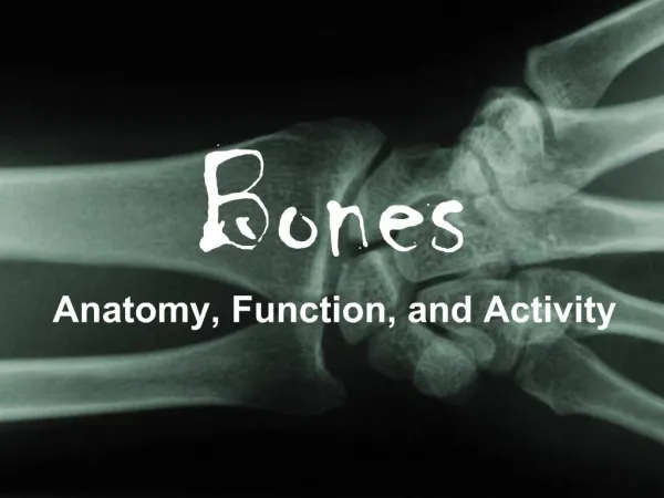
Bones. Mechanical Functions of Bones:. Protection: At numerous places inside the body bones serve to protect important and delicate organs. Ex. Brain is protected by skull Shape: Because of their rigid nature bones provide a framework around which the body is built.

E N D
Mechanical Functions of Bones: Protection: • At numerous places inside the body bones serve to protect important and delicate organs. • Ex. Brain is protected by skull Shape: • Because of their rigid nature bones provide a framework around which the body is built. • Bones are responsible for the shape and form of the human body Movement: • Working with skeletal muscles, tendons, ligaments, and joints, the bones form the moving machinery of the human body • The major role of bones in movement is that they act as levers, which make use of the forces generated by skeletal muscles in a beneficial way
Synthetic Functions of Bones Synthesis of blood cells • The major synthetic role of bones is to produce blood cells • The bones themselves are not capable of doing this. Instead, they house the bone marrow, which contains hematopoietic stem cells, capable of producing blood cells • In infants, bone marrow of all long bones is capable of synthesis, however, as a person gets older, the red marrow turns into yellow fatty marrow, which is no more capable of hematopoiesis • The red marrow in adults and older individuals is restricted to vertebrae and heads of tibia and femur
Metabolic Functions of Bones Mineral Storage • Bones serve as an important store house of minerals such as calcium and phosphorus Fat Storage • The yellow bone marrow of long bones act as a storage of fats Role in acid-base balance • Bone buffers the blood against excessive pH changes by absorbing or releasing alkaline salts
Frontal Bone This bone forms the forehead
2 Parietal Bones One bone from each side joins behind the frontal bone to form the sides and roof of the cranium
2 Temporal Bones There are two temporal bones in all, one on each side below the parietal bones
Occipital Bone This is a single bone that is present at the back of the cranium just behind the parietal and temporal bones
Sphenoid Bone This is a single bone that is situated at the base of the skull in front of the temporal bones and basilar part of the occipital bone
Ethmoid Bone This is a light and spongy bone that is situated in the anterior part of the base of the cranium. It lies between the two orbits at the roof of the nasal cavity separating the brain from the nasal cavity
Mandible This is the lower jawbone. It is U shaped and the largest and strongest bone of the face. The mandible consists of two halves that fuse at the mental symphysis by the age of two years Each half of the mandible has a horizontal body and a vertical ramus at the posterior end of the body
Maxilla The maxilla contributes to the roof of the oral cavity and the lateral walls and floor of the nasal cavity. It also provides for the attachment of the teeth of the upper jaw. It is actually two bones that are fused along the palatal fissure
2 Zygomatic Bone There are two zygomatic bones one on each side of the face forming the prominence of the cheek. Each bone also forms a part of the lateral side and floor of each orbit.
Palatine Bone The palatine is an L-shaped bone that is situated between the maxilla and the pterygoid process of the sphenoid. It is located behind the nasal cavity and hence, contributes to its floor and the lateral walls. Besides the nasal cavity, it also contributes to the roof of the mouth as well as to the floor of the orbit
Vomer It is a thin, flat single bone that lies along the midsagittal line. It articulates with the ethmoid, the sphenoid, the two palatine, and two maxillary bones each (that is 6 bones in all)
2 Lacrimal Bones There are two lacrimal bones in all, each lying in the frontal part of the medial wall of the orbit. These are the smallest bones of the face and each interacts with the frontal bone, the ethmoid bone, the maxilla, and the inferior nasal concha
2 Nasal Bones These are 2 oblong bones placed side to side that form the bridge of the nose
Inferior Nasal Concha These are paired bones of the face that arise from the maxillary bone and continues horizontally along the lateral wall of the nasal cavity. (Ahead of these bones are the middle and the superior nasal conche that are considered to be part of the cranium)
Vertebral Column The vertebral column or the spinal column is made of 33 small bones each of which is known as a vertebra. These vertebrae gives the vertebral column its flexibility due to which we can bend forwards and sideways
Cervical Vertebrate The cervical vertebrae are the first 7 vertebrae that are also the smallest of the true vertebrae. They are different from those in thoracic and lumbar regions in the fact that they have a hole or foramen in each transverse process for the vertebral process to pass through. The skull is supported on the first cervical vertebra which is known as the atlas. The second cervical vertebra is the axis which forms the pivot on which the atlas carrying the skull turns. The cervical vertebrae forms the neck
Thoracic Vertrbrae There are 12 vertebrae in all the thoracic region that come right after the cervical vertebrae. These are larger than the cervical vertebrae but smaller than those in the lumbar region. The distinct features of these vertebrae are the presence of the facets that provide the attachment of ribs Each thoracic vertebra has facets on the side of the bodies where heads of the ribs attach Other than the last two vertebrae, all the rest of the ten vertebrae have one facet on each of the transverse process for articulation with tubercles of the ribs
Lumbar Vertebrae The lumbar vertebrae consists of 5 vertebrae. They lack the foramen on transverse process that the cervical vertebrae have and the facets on the body that characterize the thoracic vertebrae
Sacral Vertebrae 5 sacral vertebrae fuse to form a triangular bone called the sacrum in adults. The sacrum fits between the two hip bones and joins the spine and the pelvis together
Coccyx The coccyx consists of 4 bones that fuse together as one grows up Coccyx can variably consist of 5 or 3 bones as well
Shoulder The shoulder is made of two bones that together allow the attachment of the arm to the body Made up from the scapula and the clavicle
Scapula The scapula is a flat triangular bone that forms the posterior part of the shoulder girdle. It connects the humerus (upper arm) with the clavicle. It is what we commonly refer to as the shoulder blade that provides for attachment of the humerus There are two shoulder blades
Clavicle It is commonly called the collarbone. It is a pair of small curved bones that join the scapula and hence, the humerus to the sternum. Together the clavicle and the scapula form the shoulder girldle
Thorax The thorax is the portion below the neck that encloses the heart and lungs
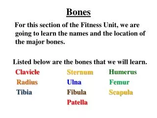
Sternum This is a long T-shaped bone. It lies in the front, central portion of the rib cage or the chest and provides for the ribs and the clavicle to attach. It consists of three parts; the topmost part is the manubrium to which the clavicle attaches, followed by the body (to which the ribs attach), and the end portion of the sternum is the xyphoid process.
Ribs (12 pairs in all) The first seven pairs are directly attached to the sternum through cartilage. The next three are attached to the sternum through a common cartilaginous extension. The last two pairs are known as the floating ribs because although they start from the thoracic vertebrae, they don not attrach to the sternum
Hip Bone This is an irregularly shaped bone that is constricted in the middle and flares at both ends. There are two hip bones that join together that along with the sacrum and the coccyx form the pelvis. Each hip bone has three parts: the ilium (flared, fan-shaped superior portion of the hip bone), ischium (lowest portion of the hip bone that curves forward and meets the pubis to form the obturator foramen) , and the pubis.
Hands The bones of the hands can be divided into those that make up the upper arm, the lower arm, the wrist, the palm and the fingers
Humerus This is a single long bone of the upper arm. It runs from the shoulder to the elbow. The humerus connects the scapula to the bones of the forearm
Radius Radius is one of the bones of the forearm that lies on the lateral side of the ulna (the other bone of the forearm). It starts from the lateral side of the elbow and continues to the thumb side of the wrist
Ulna This bone runs parallel to the radius along the forearm. It lies on the medial side of the body
Carpal Bones These are the bones of the wrist. There are 7 carpal bones in each wrist which include: • Scaphoid bones • Lunate bones • Triquetral bone • Pisiform bone • Trapezium • Trapezoid bone • Capitate bone • Hamate Bone
Metacarpal Bones These are the bones of the palms and there are 5 metacarpal bones in every palm; one corresponding to each digit
Phalanges These are the bones of the fingers. There are 5 proximal phalanges in each hand that lie in front of the metacarpals. There are 4 intermediate phalanges in front of the proximal phalanges, one on each finger other than the thumb. The last phalanges at the tip of each finger are known as the distal phalanges. There is 5.
Legs The bones of the legs can be studied as those that make up the thigh, the lower half of the legs, and the feet
Femur This is the longest bone in the human body. It is also known as the thigh bone. The head forms the ball and socket joint at the pelvic girdle.
Tibia The tibia is the second longest bone in the human body. Along with the fibula, it forms the lower part of the leg below the knee. It articulates with the femur at its superior end and with the talus at it inferior end. Laterally, it articulates with the fibula. The tibia is considered by many to be the strongest bone of the body. It is commonly known as the shin bone.
Fibula The fibula is a long but thin bone that along with the tibia forms the lower part of the human leg. It is attached to the tibia at both the ends. Its upper end articulates with the tibia at the back of its head whereas while attaching to the tibia with its lower end, it angles slightly forward. The fibula is also known as the calf bone.
Patella One on each leg, the patella is the knee cap. It is a triangular bone that forms a protective cap over the knee joint
Tarsal Bones There are 7 tarsal bones in all that lie between the tibia-fibula and the matatarsals. They are: • Calneus (heel bone) • Talus • Navicular bone • Medial cuneiform bone • Intermediate cuneiform bone • Lateral cuneiform bone • Cuboid bone
Metatarsal Bones There are 5 metatarsal bones in each foot, one corresponding to each digit. These lie between the tarsal bones and the phalanges. These may be considered to be equivalent to the metacarpal bones of the hands.
Phalanges They are the bones of the toes of the feet. There are 5 proximal phalanges in each foot that start from the metatarsals. There are 4 intermediate phalanges, one on each finger other than the big toe. The last phalanges at which each toe ends are known as the distal phalanges. They are 5 in number.
These are the main bones present in the human body. Other than these, there are 3 small bones in each of the middle ear. They are the malleus, incus and stapes. Our throat also has a small bone - the hyoid bone. These are the bones in the human body that provide for various functions. As already said, they mainly provide structural support and protection to softer organs. However, the bones of the middle ear help in conduction of sound.