Download
1 / 72
750 likes | 1.57k Vues
Muscle Overview. The three types of muscle tissue are skeletal, cardiac, and smooth These types differ in structure, location, function, and means of activation. Muscle Similarities. Skeletal and smooth muscle cells are elongated and are called muscle fibers

E N D
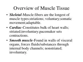
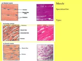
Muscle Overview • The three types of muscle tissue are skeletal, cardiac, and smooth • These types differ in structure, location, function, and means of activation
Muscle Similarities • Skeletal and smooth muscle cells are elongated and are called muscle fibers • Muscle contraction depends on two kinds of myofilaments – actin and myosin • Muscle terminology is similar • Sarcolemma – muscle plasma membrane • Sarcoplasm – cytoplasm of a muscle cell • Prefixes – myo, mys, and sarco all refer to muscle
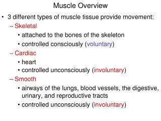
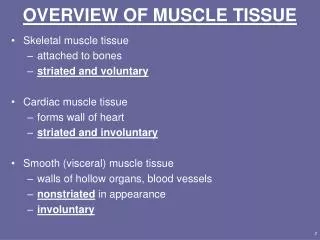
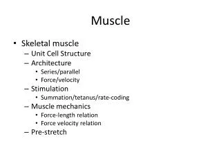
Skeletal Muscle Tissue • Packaged in skeletal muscles that attach to and cover the bony skeleton • Has obvious stripes called striations • Is controlled voluntarily (i.e., by conscious control) • Contracts rapidly but tires easily • Is responsible for overall body motility • Is extremely adaptable and can exert forces ranging from a fraction of an ounce to over 70 pounds
Cardiac Muscle Tissue • Occurs only in the heart • Is striated like skeletal muscle but is not voluntary • Contracts at a fairly steady rate set by the heart’s pacemaker • Neural controls allow the heart to respond to changes in bodily needs
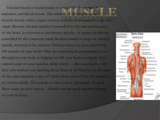
Smooth Muscle Tissue • Found in the walls of hollow visceral organs, such as the stomach, urinary bladder, and respiratory passages • Forces food and other substances through internal body channels • It is not striated and is involuntary
Muscle Function • Skeletal muscles are responsible for all locomotion • Cardiac muscle is responsible for coursing the blood through the body • Smooth muscle helps maintain blood pressure, and squeezes or propels substances (i.e., food, feces) through organs • Muscles also maintain posture, stabilize joints, and generate heat
Skeletal Muscle • Each muscle is a discrete organ composed of muscle tissue, blood vessels, nerve fibers, and connective tissue • The three connective tissue sheaths are: • Endomysium – fine sheath of connective tissue composed of reticular fibers surrounding each muscle fiber • Perimysium – fibrous connective tissue that surrounds groups of muscle fibers called fascicles • Epimysium – an overcoat of dense regular connective tissue that surrounds the entire muscle
Skeletal Muscle: Nerve and Blood Supply • Each muscle is served by one nerve, an artery, and one or more veins • Each skeletal muscle fiber is supplied with a nerve ending that controls contraction • Contracting fibers require continuous delivery of oxygen and nutrients via arteries • Wastes must be removed via veins
Nerve Stimulus of Skeletal Muscle • Skeletal muscles are stimulated by motor neurons of the somatic nervous system • Axons of these neurons travel in nerves to muscle cells • Axons of motor neurons branch profusely as they enter muscles • Each axonal branch forms a neuromuscular junction with a single muscle fiber
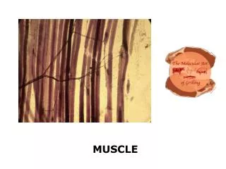
Myofibrils • Myofibrils are densely packed, rodlike contractile elements • They make up most of the muscle volume • The arrangement of myofibrils within a fiber is such that a perfectly aligned repeating series of dark A bands and light I bands is evident
Sarcomeres • The smallest contractile unit of a muscle • The region of a myofibril between two successive Z discs • Composed of myofilaments made up of contractile proteins • Myofilaments are of two types – thick and thin
Myofilaments: Banding Pattern • Thick filaments – extend the entire length of an A band • Thin filaments – extend across the I band and partway into the A band • Z-disc – coin-shaped sheet of proteins (connectins) that anchors the thin filaments and connects myofibrils to one another
Arrangement of the Filaments in a Sarcomere • Longitudinal section within one sarcomere
Role of Ionic Calcium (Ca2+) in the Contraction Mechanism • At low intracellular Ca2+ concentration: • Tropomyosin blocks the binding sites on actin • Myosin cross bridges cannot attach to binding sites on actin • The relaxed state of the muscle is enforced
Role of Ionic Calcium (Ca2+) in the Contraction Mechanism • At higher intracellular Ca2+ concentrations: • Additional calcium binds to troponin (inactive troponin binds two Ca2+) • Calcium-activated troponin binds an additional two Ca2+ at a separate regulatory site
Role of Ionic Calcium (Ca2+) in the Contraction Mechanism • Calcium-activated troponin undergoes a conformational change • This change moves tropomyosin away from actin’s binding sites
Role of Ionic Calcium (Ca2+) in the Contraction Mechanism • Myosin head can now bind and cycle • This permits contraction (sliding of the thin filaments by the myosin cross bridges) to begin
Sequential Events of Contraction Myosin head (high-energy configuration) Myosin cross bridge attaches to the actin myofilament 1 Thin filament ADP and Pi (inorganic phosphate) released Thick filament Working stroke—the myosin head pivots and bends as it pulls on the actin filament, sliding it toward the M line As ATP is split into ADP and Pi, cocking of the myosin head occurs 2 4 Myosin head (low-energy configuration) As new ATP attaches to the myosin head, the cross bridge detaches 3
Sliding Filament Model of Contraction • Thin filaments slide past the thick ones so that the actin and myosin filaments overlap to a greater degree • In the relaxed state, thin and thick filaments overlap only slightly • Upon stimulation, myosin heads bind to actin and sliding begins
Sliding Filament Model of Contraction • Each myosin head binds and detaches several times during contraction, acting like a ratchet to generate tension and propel the thin filaments to the center of the sarcomere • As this event occurs throughout the sarcomeres, the muscle shortens
Skeletal Muscle Contraction • In order to contract, a skeletal muscle must: • Be stimulated by a nerve ending • Propagate an electrical current, or action potential, along its sarcolemma • Have a rise in intracellular Ca2+ levels, the final trigger for contraction • Linking the electrical signal to the contraction is excitation-contraction coupling
Excitation-Contraction Coupling • Once generated, the action potential: • Is propagated along the sarcolemma • Travels down the T tubules • Triggers Ca2+ release from SR • Ca2+ binds to troponin and causes: • The blocking action of tropomyosin to cease • Actin active binding sites to be exposed
Excitation-Contraction Coupling • Myosin cross bridges alternately attach and detach • Thin filaments move toward the center of the sarcomere • Hydrolysis of ATP powers this cycling process • Ca2+ is removed into the SR, tropomyosin blockage is restored, and the muscle fiber relaxes
Role of Ionic Calcium (Ca2+) in the Contraction Mechanism • At low intracellular Ca2+ concentration: • Tropomyosin blocks the binding sites on actin • Myosin cross bridges cannot attach to binding sites on actin • The relaxed state of the muscle is enforced
Role of Ionic Calcium (Ca2+) in the Contraction Mechanism • At higher intracellular Ca2+ concentrations: • Additional calcium binds to troponin (inactive troponin binds two Ca2+) • Calcium-activated troponin binds an additional two Ca2+ at a separate regulatory site
Role of Ionic Calcium (Ca2+) in the Contraction Mechanism • Calcium-activated troponin undergoes a conformational change • This change moves tropomyosin away from actin’s binding sites
Role of Ionic Calcium (Ca2+) in the Contraction Mechanism • Myosin head can now bind and cycle • This permits contraction (sliding of the thin filaments by the myosin cross bridges) to begin
Sequential Events of Contraction • Cross bridge formation – myosin cross bridge attaches to actin filament • Working (power) stroke – myosin head pivots and pulls actin filament • Cross bridge detachment – ATP attaches to myosin head and the cross bridge detaches • “Cocking” of the myosin head – energy from hydrolysis of ATP cocks the myosin head into the high-energy state
Sequential Events of Contraction Myosin head (high-energy configuration) Myosin cross bridge attaches to the actin myofilament 1 Thin filament ADP and Pi (inorganic phosphate) released Thick filament Working stroke—the myosin head pivots and bends as it pulls on the actin filament, sliding it toward the M line As ATP is split into ADP and Pi, cocking of the myosin head occurs 2 4 Myosin head (low-energy configuration) As new ATP attaches to the myosin head, the cross bridge detaches 3
Functional Characteristics of Muscle Tissue • Excitability, or irritability – the ability to receive and respond to stimuli • Contractility – the ability to shorten forcibly • Extensibility – the ability to be stretched or extended • Elasticity – the ability to recoil and resume the original resting length
Muscle Twitch • A muscle twitch is the response of a muscle to a single, brief threshold stimulus • The three phases of a muscle twitch are: • Latent period – first few milli-seconds after stimulation when excitation-contraction coupling is taking place
Graded Muscle Responses • Graded muscle responses are: • Variations in the degree of muscle contraction • Required for proper control of skeletal movement • Responses are graded by: • Changing the frequency of stimulation • Changing the strength of the stimulus
Muscle Response to Varying Stimuli • A single stimulus results in a single contractile response – a muscle twitch • Frequently delivered stimuli (muscle does not have time to completely relax) increases contractile force – wave summation
Muscle Response to Varying Stimuli • More rapidly delivered stimuli result in incomplete tetanus • If stimuli are given quickly enough, complete tetanus results
Treppe: The Staircase Effect • Staircase – increased contraction in response to multiple stimuli of the same strength • Contractions increase because: • There is increasing availability of Ca2+ in the sarcoplasm • Muscle enzyme systems become more efficient because heat is increased as muscle contracts
Muscle Fatigue • Muscle fatigue – the muscle is in a state of physiological inability to contract • Muscle fatigue occurs when: • ATP production fails to keep pace with ATP use • There is a relative deficit of ATP, causing contractures • Lactic acid accumulates in the muscle • Ionic imbalances are present (extracellularpotassium) • Calcium deficiency • Neurotransmitter (ACH) rundown
Muscle Tone • Is the constant, slightly contracted state of all muscles, which does not produce active movements • Keeps the muscles firm, healthy, and ready to respond to stimulus • Essential for maintaining posture (head upright) • Essential for maintaining blood pressure (tone of smooth muscles in walls of blood vessels) • Responding to activation of stretch receptors in muscles and tendons
Muscle Metabolism: Energy for Contraction • ATP is the only source used directly for contractile activity • As soon as available stores of ATP are hydrolyzed (4-6 seconds), they are regenerated by: • The interaction of ADP with creatine phosphate (CP) • Anaerobic glycolysis • Aerobic respiration
Muscle Metabolism: Anaerobic Glycolysis • When muscle contractile activity reaches 70% of maximum: • Bulging muscles compress blood vessels • Oxygen delivery is impaired • Pyruvic acid is converted into lactic acid