Download
1 / 1
10 likes | 139 Vues
Data are expressed as mean ± SD Ki-67:Method of staining. Proliferative Activity of Mucosal and Muscular Layers in Mouse Oviduct Following Administration of Exogenous Melatonin Seyedsaadat S.N 1 ; Khajehjahromi S 1 ; Homafar M.A 1 ; Hajizadehfallah H 2 ; Mohammadghasemi F 3.

E N D
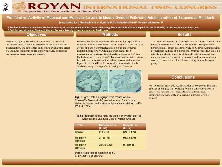
Data are expressed as mean ± SDKi-67:Method of staining Proliferative Activity of Mucosal and Muscular Layers in Mouse Oviduct Following Administration of Exogenous Melatonin Seyedsaadat S.N1; Khajehjahromi S1; Homafar M.A1; Hajizadehfallah H2; Mohammadghasemi F3 1.Student Research Committee, Guilan University of medical science, Rasht, Iran 2.Pathology Department, Poursina Hospital, Guilan University of medical science, Rasht,Iran 3.Cellular and Molecular Research Center, Guilan university of medical science, Rasht, Iran Objectives Methods Results Melatonin, a pineal hormone, is considered as a powerful antioxidant agent. It could be effective on cell cycle and cell differentiation. The aim of this study was to evaluate the effect of exogenous melatonin on proliferative activity of mucosal and muscular layers in mouse oviduct. Female adult NMRI mice were divided into 3 groups. Animals in control were received ethanol /saline and the other animals in groups of 2 and 3 were treated with 5mg/kg and 10mg/kg melatonin respectively. All animal were treated for 5 consecutive days intraperitoneally. After autopsy on 15th day, Evaluations were made by ki-67 Immunohistochemistry staining for proliferative activity of the cells in mucosal and muscular layers of tubes and Elisa for assay of serum estradiol levels. Statistical analysis was performed using ANOVA test. The mean numbers of Ki-67 positive cells in mucosal and muscular layers in controls were 2 ±2.08 and 0.96±1.10 respectively. Serum estradiol levels in controls were 80.53ng/dl. Administration of melatonin in doses of 5 mg/kg and 10mg/kg for 5 days could not alter the proliferative activity of the cells both in mucosal and muscular layers of oviduct in groups of 2 and 3 compared with controls. Serum estradiol levels were not significant between groups. Conclusions On the basis of this study, Administration of exogenous melatonin in doses of 5 mg/kg and 10 mg/kg for the 5 consecutive days in adult female mouse is not associated with alterations in proliferative activity of the mucosal and muscular layers of oviduct. Fig.1: Light Photomicrograph from mouse oviduct. Control(C), Melatonin(M) treated mouse. Dark brown stains indicates proliferative activity of cells stained by Ki-67 & H. 100X. Table1:Effect of Exogenous Melatonin on Proliferation of Mucosal and Muscular Cells in Mouse Oviduct